Hydathode pit development in the alpine plant Saxifraga cochlearis
by Wightman R., Wallis S., Aston P. (2017)
Raymond Wightman, a Simon Wallis, b Paul Aston, b
- a
- Microscopy Core Facility, Sainsbury Laboratory, University of Cambridge, Bateman Street, Cambridge, CB2 1LR, UK
- b
- Cambridge University Botanic Garden, 1 Brookside, Cambridge, CB2 1JE, UK
===
in Flora 233: 99-108 – https://doi.org/10.1016/j.flora.2017.05.018 –
https://www.sciencedirect.com/science/article/pii/S0367253017332309
Highlights
- •
-
Hydathode morphology and development were determined in Saxifraga cochlearis.
- •
-
Hydathode pits occur as an incremental developmental series along the leaf margin.
- •
-
Initiation of the pits arise through conserved divisions linked to leaf lobe maturation.
- •
-
Guard-type cells differentiate and create a thickened rim encircling the hydathode pore.
- •
-
S. cochlearis has distinct differences in epithem cell morphology compared to other types of hydathodes.
Abstract
The genus Saxifraga contain many species that form a calcified crust on the leaf surface, originating from pore-containing pits that form part of the leaf hydathode structure. The detailed morphology and development of the hydathodes are not well understood for this genus.
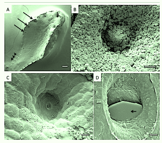
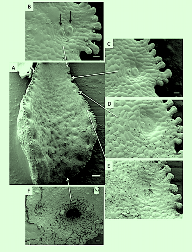
We present a study of the fine structure and developmental stages of hydathode pit formation along the leaf margin of the alpine plant Saxifraga cochlearis and cryo-fracture to reveal the internal hydathode structure. Raman- and stereo-microscopy have been used to deduce the composition and distribution of the crust.
We find the pits occur as a developmental series along the leaf where conserved and oriented divisions within leaf lobes appear to give rise to the early pit. Both pit formation and lobe maturation are linked.
As the pits deepen, hydathode pores differentiate to thick-walled, cone shaped structures and, together with the ovoid epithem tissue extrude liquid resulting in deposits of calcite that fill the pits and spill on to the leaf margin.
The epithem does not possess the typical organisation or cell morphologies that have been reported for hydathodes from other plants, lacking lobed cells and having an indistinctive sheath-like cell layer.
