Identification of potential orthologs of the key toolbox genes in a hornwort, further supporting a single ancient genetic origin of stomata in the ancestor to all stomatous land plants

==================

鉴定角苔植物中关键工具基因的潜在同源基因,进一步支持了所有有气孔陆地植物的祖先具有单一古老遗传起源的观点。

Identificação de potenciais ortólogos dos genes-chave da caixa de ferramentas em uma antócera, reforçando ainda mais a única origem genética antiga das estômatas no ancestral de todas as plantas terrestres estomáticas.

Identificación de posibles ortólogos de los genes clave de la caja de herramientas en un hornwort, respaldando aún más el origen genético antiguo único de los estomas en el ancestro de todas las plantas terrestres estomáticas.

==============

Origins and Evolution of Stomatal Development

Chater C. C. C., Caine R. S., Fleming A. J., Gray J. E. (2017)

Caspar C C Chater 123Robert S Caine 456Andrew J Fleming 456Julie E Gray 456

  • 1 Departamento de Biología Molecular de Plantas, Instituto de Biotecnología, Universidad Nacional Autónoma de Mexico, Cuernavaca 62210, Mexico (C.C.C.C.);
  • 2 Department of Molecular Biology and Biotechnology, University of Sheffield, Sheffield S10 2TN, United Kingdom (R.S.C., J.E.G.);
  • 3 Department of Animal and Plant Sciences, University of Sheffield, Sheffield S10 2TN, United Kingdom (A.J.F.)
  • 4 Departamento de Biología Molecular de Plantas, Instituto de Biotecnología, Universidad Nacional Autónoma de Mexico, Cuernavaca 62210, Mexico (C.C.C.C.).
  • 5 Department of Molecular Biology and Biotechnology, University of Sheffield, Sheffield S10 2TN, United Kingdom (R.S.C., J.E.G.);
  • 6 Department of Animal and Plant Sciences, University of Sheffield, Sheffield S10 2TN, United Kingdom (A.J.F.).

===

Plant Physiol. 2017 Jun;174(2):624-638 – doi: 10.1104/pp.17.00183 – Epub 2017 Mar 29 – PMID: 28356502 – PMCID: PMC5462063 –

https://pubmed.ncbi.nlm.nih.gov/28356502/

Abstract

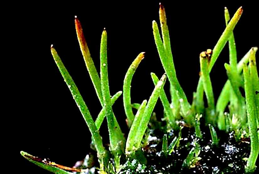
The fossil record suggests stomata-like pores were present on the surfaces of land plants over 400 million years ago. Whether stomata arose once or whether they arose independently across newly evolving land plant lineages has long been a matter of debate. In Arabidopsis, a genetic toolbox has been identified that tightly controls stomatal development and patterning. This includes the basic helix-loop-helix (bHLH) transcription factors SPEECHLESS (SPCH), MUTEFAMA, and ICE/SCREAMs (SCRMs), which promote stomatal formation. These factors are regulated via a signaling cascade, which includes mobile EPIDERMAL PATTERNING FACTOR (EPF) peptides to enforce stomatal spacing. Mosses and hornworts, the most ancient extant lineages to possess stomata, possess orthologs of these Arabidopsis (Arabidopsis thaliana) stomatal toolbox genes, and manipulation in the model bryophyte Physcomitrella patens has shown that the bHLH and EPF components are also required for moss stomatal development and patterning. This supports an ancient and tightly conserved genetic origin of stomata. Here, we review recent discoveries and, by interrogating newly available plant genomes, we advance the story of stomatal development and patterning across land plant evolution. Furthermore, we identify potential orthologs of the key toolbox genes in a hornwort, further supporting a single ancient genetic origin of stomata in the ancestor to all stomatous land plants.

Stomata in hornworts are primarily involved in sporophyte desiccation and spore discharge rather than the regulation of photosynthesis-related gaseous exchange

Hornwort stomata do not respond actively to exogenous and environmental cues

by Pressel S., Renzaglia K. S., Clymo R. S., Duckett, J. G. (2018)

Silvia Pressel,1Karen S Renzaglia,2Richard S (Dicky) Clymo,3 and Jeffrey G Duckett, 1

1Life Sciences Department, Natural History Museum, London, UK

2Plant Biology Department, Southern Illinois University, Carbondale, USA

3School of Biological and Chemical Sciences, Queen Mary University of London, London, UK

===

In Annals of Botany 122(1): 45–57 – https://doi.org/10.1093/aob/mcy045

https://www.ncbi.nlm.nih.gov/pmc/articles/PMC6025193/

Abstract

Backgrounds and Aims

Because stomata in bryophytes occur on sporangia, they are subject to different developmental and evolutionary constraints from those on leaves of tracheophytes. No conclusive experimental evidence exists on the responses of hornwort stomata to exogenous stimulation.

Methods

Responses of hornwort stomata to abscisic acid (ABA), desiccation, darkness and plasmolysis were compared with those in tracheophyte leaves. Potassium ion concentrations in the guard cells and adjacent cells were analysed by X-ray microanalysis, and the ontogeny of the sporophytic intercellular spaces was compared with those of tracheophytes by cryo-scanning electron microscopy.

Key Results

The apertures in hornwort stomata open early in development and thereafter remain open. In hornworts, the experimental treatments, based on measurements of >9000 stomata, produced only a slight reduction in aperture dimensions after desiccation and plasmolysis, and no changes following ABA treatments and darkness. In tracheophytes, all these treatments resulted in complete stomatal closure. Potassium concentrations are similar in hornwort guard cells and epidermal cells under all treatments at all times. The small changes in hornwort stomatal dimensions in response to desiccation and plasmolysis are probably mechanical and/or stress responses of all the epidermal and spongy chlorophyllose cells, affecting the guard cells. In contrast to their nascent gas-filled counterparts across tracheophytes, sporophytic intercellular spaces in hornworts are initially liquid filled.

Conclusions

Our experiments demonstrate a lack of physiological regulation of opening and closing of stomata in hornworts compared with tracheophytes, and support accumulating developmental and structural evidence that stomata in hornworts are primarily involved in sporophyte desiccation and spore discharge rather than the regulation of photosynthesis-related gaseous exchange. Our results run counter to the notion of the early acquisition of active control of stomatal movements in bryophytes as proposed from previous experiments on mosses.


Stomata in early land plants

Vegetative and reproductive innovations of early land plants: implications for a unified phylogeny

by Renzaglia K. S., Duff R. J., Nickrent D. L., Garbary D. J. (2000)

Karen Sue RenzagliaR. Joel DuffDaniel L. Nickrent, David J. Garbary,

In Philosoph. Transactions Roy. Soc. B, Biol. Sci. – https://doi.org/10.1098/rstb.2000.0615

https://royalsocietypublishing.org/doi/10.1098/rstb.2000.0615

Abstract

As the oldest extant lineages of land plants, bryophytes provide a living laboratory in which to evaluate morphological adaptations associated with early land existence. In this paper we examine reproductive and structural innovations in the gametophyte and sporophyte generations of hornworts, liverworts, mosses and basal pteridophytes. Reproductive features relating to spermatogenesis and the architecture of motile male gametes are overviewed and evaluated from an evolutionary perspective. Phylogenetic analyses of a data set derived from spermatogenesis and one derived from comprehensive morphogenetic data are compared with a molecular analysis of nuclear and mitochondrial small subunit rDNA sequences.

Although relatively small because of a reliance on water for sexual reproduction, gametophytes of bryophytes are the most elaborate of those produced by any land plant. Phenotypic variability in gametophytic habit ranges from leafy to thalloid forms with the greatest diversity exhibited by hepatics. Appendages, including leaves, slime papillae and hairs, predominate in liverworts and mosses, while hornwort gametophytes are strictly thalloid with no organized external structures. Internalization of reproductive and vegetative structures within mucilage–filled spaces is an adaptive strategy exhibited by hornworts. The formative stages of gametangial development are similar in the three bryophyte groups, with the exception that in mosses apical growth is intercalated into early organogenesis, a feature echoed in moss sporophyte ontogeny.

A monosporangiate, unbranched sporophyte typifies bryophytes, but developmental and structural innovations suggest the three bryophyte groups diverged prior to elaboration of this generation. Sporophyte morphogenesis in hornworts involves non–synchronized sporogenesis and the continued elongation of the single sporangium, features unique among archegoniates. In hepatics, elongation of the sporophyte seta and archegoniophore is rapid and requires instantaneous wall expandability and hydrostatic support. Unicellular, spiralled elaters and capsule dehiscence through the formation of four regular valves are autapomorphies of liverworts. Sporophytic sophistications in the moss clade include conducting tissue, stomata, an assimilative layer and an elaborate peristome for extended spore dispersal. Characters such as stomata and conducting cells that are shared among sporophytes of mosses, hornworts and pteridophytes are interpreted as parallelisms and not homologies.

Our phylogenetic analysis of three different data sets is the most comprehensive to date and points to a single phylogenetic solution for the evolution of basal embryophytes. Hornworts are supported as the earliest divergent embryophyte clade with a moss/liverwort clade sister to tracheophytes. Among pteridophytes, lycophytes are monophyletic and an assemblage containing ferns, Equisetum and psilophytes is sister to seed plants. Congruence between morphological and molecular hypotheses indicates that these data sets are tracking the same phylogenetic signal and reinforces our phylogenetic conclusions. It appears that total evidence approaches are valuable in resolving ancient radiations such as those characterizing the evolution of early embryophytes. More information on land plant phylogeny can be found at: http://www.science.siu.edu/landplants/index.html.

The evolution of the stomatal apparatus

Figure 3.Cryo-scanning electron micrographs of freeze-fractured hornwort gametophytes (a–c) and sporophytes (d–i): Anthoceros agrestis (a,c,d–f); Folioceros fusiformis (b); Leiosporoceros dussii (g); Megaceros enigmaticus (h); Dendroceros granulatus (i). Sections through thalli showing mucilage-filled cavities (asterisk). (cNostoc colony. (d,g) Intercellular spaces are initially liquid-filled (asterisk) but become gas-filled (e, arrowed) following stomatal opening. (f) Columella with gas-filled (asterisk) intercellular spaces. (h,i) Young (h) and mature (i) sporophytes of astomate taxa, showing complete absence of intercellular spaces in the assimilatory layers which collapse and dry (i). Scale bars: (a,b) 200 µm; (d,e,g) 50 µm; (c,f,h,i) 20 µm.

The evolution of the stomatal apparatus: intercellular spaces and sporophyte water relations in bryophytes—two ignored dimensions

by Duckett J. G., Pressel S. (2017)

Jeffrey G. Duckett, Silvia Pressel,

In Philosoph. Transactions Royal Soc. B Biol. Sci. https://doi.org/10.1098/rstb.2016.0498

https://royalsocietypublishing.org/doi/full/10.1098/rstb.2016.0498

Figure 4.Cryo-scanning electron micrographs of freeze-fractured moss sporophytes: Physcomitrella patens (a,b); Physcomitrium pyriforme(c,d); Lyellia crispa (e,f). (a,c) Young sporophytes with liquid-filled (asterisk) intercellular spaces. (e) Gas (arrowed) gradually replaces their initially liquid-filled content following stomatal opening, as evidenced by the presence of intercellular spaces only partially filled with liquid (asterisk in f). Liquid is first lost from the substomatal cavities (b; S, stoma) until the entire intercellular space system becomes gas-filled (d). Scale bars: (c,d) 100 µm; (a,e) 50 µm; (b,d) 20 µm.

Abstract

Cryo-scanning electron microscopy shows that nascent intercellular spaces (ICSs) in bryophytes are liquid-filled, whereas these are gas-filled from the outset in tracheophytes except in the gametophytes of Lycopodiales.

ICSs are absent in moss gametophytes and remain liquid-filled in hornwort gametophytes and in both generations in liverworts. Liquid is replaced by gas following stomatal opening in hornworts and is ubiquitous in moss sporophytes even in astomate taxa.

Figure 5.Cryo-scanning electron micrographs of freeze-fractured moss sporophytes: Polytrichum juniperinum (a,b); Mnium hornum (c); Atrichum undulatum (d); Pogonatum aloides (e,f). (a,b) Unopened (a) and open (b) stoma subtended by a gas-filled intercellular space. (c) Sunken stoma subtended by a liquid-filled intercellular space. (d–f) In astomate taxa, intercellular spaces are also initially liquid-filled (asterisk, e) and the same process of liquid replacement by gas occurs in their fully expanded capsules (d,f). Scale bars: (f) 200 µm; (a–e) 20 µm.

New data on moss water relations and sporophyte weights indicate that the latter are homiohydric while X-ray microanalysis reveals an absence of potassium pumps in the stomatal apparatus.

The distribution of ICSs in bryophytes is strongly indicative of very ancient multiple origins. Inherent in this scenario is either the dual or triple evolution of stomata. The absence, in mosses, of any relationship between increases in sporophyte biomass and stomata numbers and absences, suggests that CO2 entry through the stomata, possible only after fluid replacement by gas in the ICSs, makes but a minor contribution to sporophyte nutrition. Save for a single claim of active regulation of aperture dimensions in mosses, all other functional and structural data point to the sporophyte desiccation, leading to spore discharge, as the primeval role of the stomatal apparatus.

This article is part of a discussion meeting issue ‘The Rhynie cherts: our earliest terrestrial ecosystem revisited’.

Hornwort stomata walls

Hornwort stomata walls are not built for movement

Assiry A. (2019)

In Botany One March 20, 2019 –

https://www.botany.one/2019/03/hornwort-stomata-walls-are-not-built-for-movement/

Guard cell walls are built to resist bending and deformation to open and close the pore. Pectins provide flexibility and resilience to walls; in particular arabinans and unesterified homo-galacturonans are required for stomata function. 

Merced and Renzaglia use immunolabelling to investigate how wall architecture and pectin composition of Arabidopsis stomata compare to the unresponsive stomata of the hornwort Phaeoceros (Notothyladaceae, Anthocerotophyta).

(Continued)

Variations in guard cell wall composition reflect different physiological activity of stomata in land plants


Contrasting pectin polymers in guard cell walls of Arabidopsis and the hornwort Phaeoceros reflect physiological differences

by Merced A., Renzaglia K. S. (2018)

Amelia Merced,  Karen S. Renzaglia,

===

in Annals of Botany, mcy168 – https://doi.org/10.1093/aob/mcy168 –

https://academic.oup.com/aob/advance-article-abstract/doi/10.1093/aob/mcy168/5092734?redirectedFrom=fulltext

Abstract

Background and Aims

In seed plants, stomata regulate CO2 acquisition and water relations via transpiration, while minimizing water loss. Walls of guard cells are strong yet flexible because they open and close the pore by changing shape over the substomatal cavity. Pectins are necessary for wall flexibility and proper stomata functioning. This study investigates the differences in pectin composition in guard cells of two taxa that represent key lineages of plants with stomata: Arabidopsis, an angiosperm with diurnal stomatal activity, and Phaeoceros, a bryophyte that lacks active stomatal movement.

Methods

Using immunolocalization techniques in transmission electron microscopy, this study describes and compares the localization of pectin molecule epitopes essential to stomata function in guard cell walls of Arabidopsis and Phaeoceros.

Key Results

In Arabidopsis, unesterified homogalacturonans very strongly localize throughout guard cell walls and are interspersed with arabinan pectins, while methyl-esterified homogalacturonans are restricted to the exterior of the wall, the ledges and the junction with adjacent epidermal cells. In contrast, arabinans are absent in Phaeoceros, and both unesterified and methyl-esterified homogalacturonans localize throughout guard cell walls.

Conclusions

Arabinans and unesterified homogalacturonans are required for wall flexibility, which is consistent with active regulation of pore opening in Arabidopsis stomata. In contrast, the lack of arabinans and high levels of methyl-esterified homogalacturonans in guard cell walls of Phaeoceros are congruent with the inability of hornwort stomata to open and close with environmental change. Comparisons across groups demonstrate that variations in guard cell wall composition reflect different physiological activity of stomata in land plants.

A lack of physiological regulation of stomatal movements in hornworts compared with tracheophytes

hornwortyoung
Introduction to the Anthocerotophyta (UCMP), Berkeley Above, you can see pictures of the hornwort Phaeoceros. On the left is a plant with young sporophytes beginning to elongate from the top of the gametophyte. (http://www.ucmp.berkeley.edu/plants/anthocerotophyta.html)


Hornwort stomata do not respond actively to exogenous and environmental cues

by Pressel S., Renzaglia K. S., Clymo R. S., Duckett, J. G. (2018) 

Silvia Pressel, Karen S. Renzaglia, Richard S. (Dicky) Clymo, Jeffrey G. Duckett


in Annals of Botany, mcy045 –  https://doi.org/10.1093/aob/mcy045

https://academic.oup.com/aob/advance-article-abstract/doi/10.1093/aob/mcy045/4979633?redirectedFrom=fulltext

Abstract

Backgrounds and Aims

Because stomata in bryophytes occur on sporangia, they are subject to different developmental and evolutionary constraints from those on leaves of tracheophytes. No conclusive experimental evidence exists on the responses of hornwort stomata to exogenous stimulation.


Methods

Responses of hornwort stomata to abscisic acid (ABA), desiccation, darkness and plasmolysis were compared with those in tracheophyte leaves. Potassium ion concentrations in the guard cells and adjacent cells were analysed by X-ray microanalysis, and the ontogeny of the sporophytic intercellular spaces was compared with those of tracheophytes by cryo-scanning electron microscopy.


Key Results

The apertures in hornwort stomata open early in development and thereafter remain open. In hornworts, the experimental treatments, based on measurements of >9000 stomata, produced only a slight reduction in aperture dimensions after desiccation and plasmolysis, and no changes following ABA treatments and darkness. In tracheophytes, all these treatments resulted in complete stomatal closure. Potassium concentrations are similar in hornwort guard cells and epidermal cells under all treatments at all times. The small changes in hornwort stomatal dimensions in response to desiccation and plasmolysis are probably mechanical and/or stress responses of all the epidermal and spongy chlorophyllose cells, affecting the guard cells. In contrast to their nascent gas-filled counterparts across tracheophytes, sporophytic intercellular spaces in hornworts are initially liquid filled.


Conclusions

Our experiments demonstrate a lack of physiological regulation of opening and closing of stomata in hornworts compared with tracheophytes, and support accumulating developmental and structural evidence that stomata in hornworts are primarily involved in sporophyte desiccation and spore discharge rather than the regulation of photosynthesis-related gaseous exchange. Our results run counter to the notion of the early acquisition of active control of stomatal movements in bryophytes as proposed from previous experiments on mosses.

==================

https://www.botany.one/2018/07/hornwort-stomata-are-not-actively-regulated/

Hornwort stomata are not actively regulated

Stomata, pores in the plant epidermis that regulate gas exchange, are a key innovation that enabled freshwater algae to colonize Earth’s landmasses some 500 Mya. Because stomata in bryophytes occur on sporangia, they are subject to different developmental and evolutionary constraints from those on leaves of tracheophytes. No conclusive experimental evidence exists on the responses of hornwort stomata to exogenous stimulation.

Hornwort stomata

Pressel and colleagues investigate stomatal behaviour in hornworts. They investigated responses of hornwort stomata to abscisic acid (ABA), desiccation, darkness and plasmolysis and compared these with those in tracheophyte leaves. Potassium ion concentrations in the guard cells and adjacent cells were analysed by X-ray microanalysis, and the ontogeny of the sporophytic intercellular spaces was compared with those of tracheophytes by cryo-scanning electron microscopy.

They show that there are no potassium fluxes associated with hornwort stomata, and that these do not respond to external factors (abscisic acid, desiccation, darkness and plasmolysis), which cause stomatal closure in other land plants. Their results run counter to the notion that active stomatal control was acquired early in the evolution of land plants, and support the alternative hypothesis of gradual acquisition of active control mechanisms.

Hornwort Stomata (Anthoceros)

Screen Shot 2018-02-07 at 21.21.43


Anatomy, Ultrastructure and Physiology of Hornwort Stomata

by Lucas J. R. (2000)

Jessica Regan Lucas,  Southern Illinois University Carbondale

========

in  Honors Theses. Paper 136 –

http://opensiuc.lib.siu.edu/cgi/viewcontent.cgi?article=1142&context=uhp_theses

Screen Shot 2018-02-07 at 21.24.07

Conclusions;

The development of hornwort stomata is very simple. This is indicated by the single longitudinal division of the guard cell precursor, pectinous ledges, lack of subsidiary cells, and lack of radial micellation. Gas exchange seems to be a likely function of hornwort stomata, but the absence of vascular tissue makes water transport improbable. Histochemical stains for malate and potassium indicate that guard cells localize ions for a short time- after the differentiation of the epidermis and before spore dispersal.

Diurnal guard cell movements do not occur in hornworts. Neither dehydration or ABA treatment effects the guard cells in respect to movement. It is still unclear whether or not hornwort stomata are homologous to stomata of vascular plants. The prominent chloroplast, the localization of ions, and the role of stomata in gas exchange suggest that anthocerote stomata are related to those of other embryophytes.

However, the lack of vascular tissue and stomatal movement counter the homologous theory. Also in opposition to this paradigm is the distinct wall structure of hornwort guard cells. A multilayered wall and ledges of pectin have only been reported for a few other plants. In the future to elucidate the homology of these structures, the effect of ABA should further be studied. Also the guard cells’ ability to transport ions, which is essential for movement, should be determined.

Structure and function of hornwort stomata.

Photo credit: Google

The hornwort Phaeoceros

Structure and function of hornwort stomata

by Lucas J. R., Renzaglia K. S. (2002)

Jessica R. Lucas, Karen S. Renzaglia, Department of Plant Biology, Southern Illinois University, Carbondale, IL 62901

in Microscopy and Microanalysis 8, Supplement 2: 1090–1091 – DOI 10.1017.S143192760210701X –

As minute structures that open and close in response to environmental cues, stomata are key adaptive innovations of land plants [1]. In vascular plants, stomata function in gas exchange in photosynthetic tissue and in facilitating nutrient transport through the transpirational stream. In bryophytes, the basal most land plants, stomata are restricted in occurrence and are poorly characterized in regards to structure-function relationships.

This study was undertaken to evaluate the microanatomy and physiology of stomata in hornworts, the oldest living lineage of plants that possess stomata. Protocols for SEM and TEM follow Renzaglia et al. [2]. To assess the osmotic regulation of stomatal functioning, we performed cytochemical localizations of potassium with Macullum’s reagent [3] and organic ions with Fast Violet B solution [4].

In hornworts, stomata occur scattered among elongated epidermal cells in the sporophyte. Mature stomata each consist of two bean-shaped guard cells subtended by an air-filled cavity (Figs. 1-3). In cross section, guard cells are larger than epidermal cells and they each contain a large starchfilled chloroplast (Figs. 1, 2).

Aside from prominent ledges on the outer and inner walls where the cells meet, the guard cell wall is thin throughout (Fig. 1). Extensive fibrous and flocculent material is associated with the outer ledge where the pore forms (Figs. 1, 3). The large chloroplasts in the underlying photosynthetic tissue are invariably positioned adjacent to air spaces, indicating a role of stomata in gas exchange (Fig. 4).

In contrast, the lack of an internal conducting system in hornworts suggests that stomata are not involved in nutrient transport or in regulating the transpirational flow as they are in plants with vascular tissue. Stomata examined in immature regions of the sporophyte are closed, while in mature regions, the stomata are typically opened.

Examination of sporophytes collected during dark and light periods indicates that there are no diurnal movements, i.e., guard cells in hornworts do not open and close in response to light. Further experiments with abscisic acid, a hormone that results in stomatal closure in vascular plants, resulted in no change in the pore (not illustrated).

In immature regions, both K+ and malate exhibit general localization in all epidermal cells (Figs. 5, 7). In mature tissue, these ions are localized in guard cells and are interpreted as functioning as osmoticum that is responsible for water influx and the creation and maintenance of stomatal pores (Figs. 6, 8).

These results coupled with the structural observations above support the concept that hornwort stomata open once through osmotic changes and remain open during the duration of physiological activity of the sporophyte. It appears that unlike vascular plants, the primary role of stomata in hornworts is in providing a passageway for gas exchange.

Architecture and fate of stomata in hornworts

Figure 4.Stages of senescence and collapse of stomata in three genera of hornworts. The outer aperture remains open and increases in diameter during the drying process. A, P. carolinianus. Scanning electron microscopy (SEM) shows newly opened, slightly raised stoma directly above the involucre. B, L. dussii. Cross section light micrograph of a newly opened stoma shows large starch-filled plastids in guard cells and differentially thickened epidermal and guard cell walls. Small plastids (arrow) occur in epidermal cells, and a substomatal cavity (asterisk) leads to intercellular spaces in the assimilative (cortical) tissue. C, A adscendens Lehm. and Lindenb. SEM cross section shows the epidermis and a stoma with dead collapsing guard cells that contain degenerated protoplasm (arrow). Adjacent epidermal cells have thickened radial walls and are beginning to collapse in the opposite direction from the guard cells. The aperture is wide open superficially, and the thin ventral guard cell walls are buckled. A large substomatal cavity (asterisk) leads to internal air spaces. D, A. adscendens. SEM of stoma shows the onset of guard cell collapse before epidermal cells dry. A thicker cuticle covers epidermal cells compared with guard cells. E, L. dussii. Cross section light micrograph shows dead guard cells with degenerated protoplasm at the onset of collapse of the outer cell wall and while fluid is still within the substomatal cavity (asterisk) and intercellular spaces (double asterisks). Small plastids (arrow) in epidermal cells contrast with large starch-filled plastids (p) in assimilative cells. F, L. dussii. TEM of dead, collapsed stoma shows the coordinated folding of the thin ventral walls of guard cells. The aperture is open from the outside due to the rigid outer ledges. G, A. adscendens. SEM shows completely collapsed guard cells surrounded by hydrated epidermal cells. Stoma diameter is greater than in the precollapsed guard cell in E. The outer aperture is open, and folded ventral walls of guard cells are visible internally (arrow). H, L. dusii. Cross section light micrograph shows collapsed guard cells. The adjacent epidermal cell contains degenerated cytoplasm and has begun to collapse like an accordion in the opposite direction from the guard cells. Assimilative cells begin to die around the substomatal cavity (asterisk) and intercellular space (double asterisks). I, P. carolinianus. SEM shows the epidermis in desiccated and dehisced sporophyte with ridges of collapsed epidermal cell surrounding an enlarged stoma that has a broadened outer aperture. Bars = 5 μm except for F, where bar = 2 μm.


Hornwort Stomata: Architecture and Fate Shared with 400-Million-Year-Old Fossil Plants without Leaves

by Renzaglia K. S., Villarreal J. C., Piatkowski B. T., Lucas J. R., Merced A. (2017)

Karen S. Renzaglia, Juan Carlos Villarreal, Bryan T. Piatkowski, Jessica R. Lucas, and Amelia Merced

Department of Plant Biology, Southern Illinois University, Carbondale, Illinois 62901-6509 (K.S.R., J.R.L.);

Département de Biologie, Université Laval, Quebec, Quebec, Canada G1V 0A6 (J.C.V.);

Smithsonian Tropical Research Institute, Ancon, 0843-03092 Panama, Republic of Panama (J.C.V.);

Department of Biology, Duke University, Durham, North Carolina 27708 (B.T.P.);

Institute of Neurobiology, University of Puerto Rico, San Juan, Puerto Rico 00901 (A.M.)


in Plant Physiology, June 2017, Vol. 174, pp. 788–797

Screen Shot 2017-06-07 at 21.07.21

Abstract

As one of the earliest plant groups to evolve stomata, hornworts are key to understanding the origin and function of stomata.

Hornwort stomata are large and scattered on sporangia that grow from their bases and release spores at their tips. We present data from development and immunocytochemistry that identify a role for hornwort stomata that is correlated with sporangial and spore maturation.

We measured guard cells across the genera with stomata to assess developmental changes in size and to analyze any correlation with genome size. Stomata form at the base of the sporophyte in the green region, where they develop differential wall thickenings, form a pore, and die.

Screen Shot 2017-06-07 at 21.09.21

Guard cells collapse inwardly, increase in surface area, and remain perched over a substomatal cavity and network of intercellular spaces that is initially fluid filled. Following pore formation, the sporophyte dries from the outside inwardly and continues to do so after guard cells die and collapse.

Spore tetrads develop in spore mother cell walls within a mucilaginous matrix, both of which progressively dry before sporophyte dehiscence.

A lack of correlation between guard cell size and DNA content, lack of arabinans in cell walls, and perpetually open pores are consistent with the inactivity of hornwort stomata.

Stomata are expendable in hornworts, as they have been lost twice in derived taxa. Guard cells and epidermal cells of hornworts show striking similarities with the earliest plant fossils.

Our findings identify an architecture and fate of stomata in hornworts that is ancient and common to plants without sporophytic leaves.