Stomata in Equisetum and Psilotum

Photo credit: AOB

Drawings of Equisetum stomata. (A) E. palustre; fig. 10 from Duval-Jouve (1864). (B) E. fluviatile; fig. 12 from Riebner (1925).

The remarkable stomata of horsetails (Equisetum): patterning, ultrastructure and development

byErin Cullen,

Paula J. Rudall.paula-rudall-cropped

(2016)

in Ann Bot (2016)doi: 10.1093/aob/mcw094 – 

http://aob.oxfordjournals.org/content/early/2016/06/03/aob.mcw094.abstract

F3.small
Equisetum myriochaetum (A, B, LM; C, DIC; E, F, SEM). (A) Thin paradermal section of a mature stoma showing radiating ribs on subsidiary cells. (B) Thick paradermal section of a mature stoma with radiating ribs. Both guard cells and superadjacent subsidiary cells are visible. (C) Oblique view of a mature stoma showing radiating ribs. (D) Transverse section of a mature sunken stoma, showing silica on the surface of subsidiary cells. (E) Macerated stoma showing radiating ribs. gc, guard cell; gcn, guard cell nucleus; rr, radiating ribs; sc, subsidiary cell; sc, silica. Scale bars: 10 μm in (A−D), 5 μm in (E). – http://aob.oxfordjournals.org/content/early/2016/06/03/aob.mcw094/F3.small.gif

Abstract

Background and Aims The stomata of Equisetum – the sole extant representative of an ancient group of land plants – are unique with respect to both structure and development, yet little is known about details of ultrastructure and patterning, and existing accounts of key developmental stages are conflicting.

Methods We used light and electron microscopy to examine mature stomata and stomatal development in Equisetum myriochaetum, and compared them with other land plants, including another putative fern relative, Psilotum. We reviewed published reports of stomatal development to provide a comprehensive discussion of stomata in more distantly related taxa.

Screen Shot 2018-04-18 at 20.05.43
Equisetum myriochaetum (TEM). (A, B) Transverse sections of mature stomata. (C) Paradermal view of a mature stomatal complex with radiating ribs; parts of both guard cells and superadjacent subsidiary cells are visible in this plane of the section, which lies below the outer ledges that delimit the pore. (D) Detail of interlocking outer cuticular ledges on subsidiary cells, and thinner ledges on guard cells below. (E) Transverse section of a young stoma. (F) Transverse section of a mesophyll cell below the stoma. chl, chloroplast; gc, guard cell; icl, inner cuticular ledge (on guard cells); ocl, outer cuticular ledge (on subsidiary cells); rr, radiating ribs; sc, subsidiary cell. Scale bars: 10 μm in (A–C), 2 μm in (D−F).

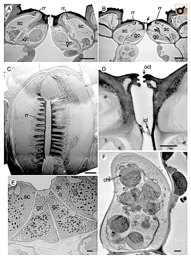
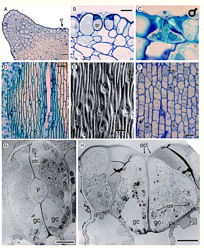
Key Results Stomatal development in Equisetum is basipetal and sequential in strict linear cell files, in contrast with Psilotum, in which stomatal development occurs acropetally. In Equisetum, cell asymmetry occurs in the axial stomatal cell file, resulting in a meristemoidal mother cell that subsequently undergoes two successive asymmetric mitoses. Each stomatal cell complex is formed from a single precursor meristemoid, and consists of four cells: two guard cells and two mesogene subsidiary cells. Late periclinal divisions occur in the developing intervening cells.

Screen Shot 2018-04-18 at 20.07.51
Equisetum myriochaetum, stomatal development (A, C−F, LM; B, SEM; all images oriented with plant apex uppermost). (A) Composite image showing the series of developmental stages along a single axial stomatal cell file. (B) Series of developmental stages in surface view, increasingly sunken towards the apex. (C) Longitudinal section of a stem with fully differentiated stomata arrowed; less well-developed stomata are closer to the internode. (D) Undifferentiated cells in a stomatal cell file, close to the internode; meristemoids are slightly larger than intervening cells. (E) Later stages of development, showing initial asymmetric cell division and the resulting pair of cells. (F) Later stages of development, showing the second asymmetric cell division and resulting triad. (G) Differentiated stomatal complex. gc, guard cell; gmc, guard mother cell; ic, intervening cell; m, meristemoid; sc, subsidiary cell, st, stoma. Scale bars = 20 μm in (A), 100 μm in (B), (C), 7.5 μm in (D), (F), (G).

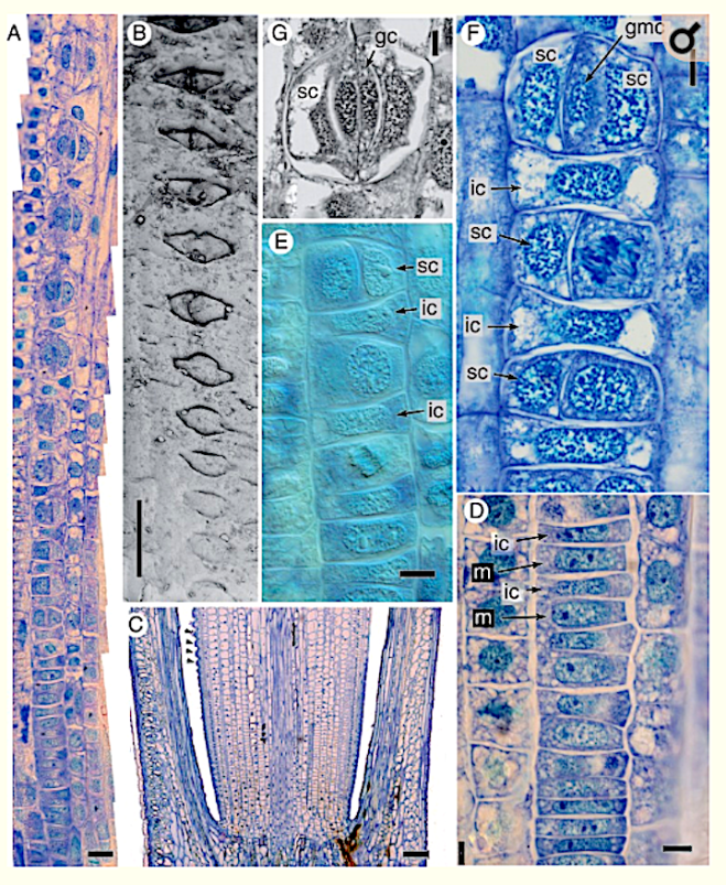
Conclusions In addition to the unique mature structure, several highly unusual developmental features include a well-defined series of asymmetric and symmetric mitoses in Equisetum, which differs markedly from Psilotum and other land plants. The results contribute to our understanding of the diverse patterns of stomatal development in land plants, including contrasting pathways to paracytic stomata. They add to a considerable catalogue of highly unusual traits of horsetails – one of the most evolutionarily isolated land-plant taxa.

Screen Shot 2018-04-18 at 20.10.12
Stomata of Psilotum (A, B, E, F, G, H, P. nudum; C, D, P. intermedium). (A, B) P. nudum, transverse section of a mature stem with detail of a stoma in (B) (C) P. intermedium, transverse section of a mature stoma. (D) P. intermedium, LM stem surface. (E) P. nudum, SEM stem surface. (F) P. nudum, paradermal section of the epidermis with guard mother cells and a recently divided stoma. (G) P. nudum, TEM paradermal section of a mature stoma. (G) P. nudum, TEM transverse section of a mature stoma (slightly off-centre, since most stomata are not quite parallel with the axis). chl, chloroplast; gc, guard cell; m, meristemoid; st, stoma. Scale bars = 50 μm in (A), 10 μm in (B), (C), (G), (H); 100 μm in (D), (E); 25 μm in (F).

AHA1 plays a major role in blue light-dependent stomatal opening


Plasma membrane H+-ATPase1 (AHA1) plays a major role in Arabidopsis thaliana for stomatal opening in response to blue light

by Yamauchi S., Takemiya A., Sakamoto T., Shimazaki K.-i. (2016)

in Plant physiology · June 2016 –

https://www.researchgate.net/publication/303797064_Plasma_membrane_H-ATPase1_AHA1_plays_a_major_role_in_Arabidopsis_thaliana_for_stomatal_opening_in_response_to_blue_light 

Abstract
Stomata open in response to a weak blue light under strong red light illumination. A blue light signal is perceived by phototropins and transmitted to the plasma membrane H+-ATPase that drivesstomatal opening.
To identify the components in this pathway, we screened for mutants impaired in blue light-dependent stomatal opening. We analyzed one such mutant, provisionally named blus2 (blue light signaling2), and found that stomatal opening in leaves was impaired by 65%, although the magnitude of red light-induced opening was not affected.
Blue light-dependent stomatal opening in the epidermis and H+ pumping in guard cell protoplasts were inhibited by 70% in blus2. Whole genome re-sequencing identified a mutation in the AHA1 gene of the mutant at Gly-602. T-DNA insertion mutants of AHA1 exhibited a similar phenotype to blus2; this phenotype was complemented by the AHA1 gene.
We renamed blus2 as aha1-10. T-DNA insertion mutants of AHA2 and AHA5 did not show any impairment in stomatal response, although the transcript levels of AHA2 and AHA5 were higher than those of AHA1 in wild-type guard cells.
Stomata in ost2, a constitutively active AHA1 mutant, did not respond to blue light. A decreased amount of H+-ATPase in aha1-10 accounted for the reduced stomatal blue light responses and the decrease was likely caused by proteolysis of misfolded AHA1.
From these results, we conclude that AHA1 plays a major role in blue light-dependent stomatal opening in Arabidopsis and that the mutation made the AHA1 protein unstable in guard cells.

Cytosolic Ca2+ in guard cells and blue light


Measurement of changes in cytosolic Ca2+ in Arabidopsis guard cells and mesophyll cells in response to blue light.

by Harada A., Shimazaki K. (2009)

in Plant Cell Physiol.50, 360–373. doi: 10.1093/pcp/pcn203 –

PubMed Abstract | CrossRef Full Text

http://pcp.oxfordjournals.org/content/50/2/360

Abstract

Phototropins (phot1 and phot2) are blue light (BL) receptors that mediate responses including phototropism, chloroplast movement and stomatal opening, and increased cytosolic Ca2+.

BL absorbed by phototropins activates plasma membrane H+-ATPase in guard cells, resulting in membrane hyperpolarization, and drives K+ uptake and stomatal opening. However, it is unclear whether the phototropin-mediated Ca2+ increase activates the H+-ATPase.

Here, we determined cytosolic Ca2+concentrations in guard cell protoplasts (GCPs) from Arabidopsis transformed with aequorin. Cytosolic Ca2+ increased rapidly in response to BL in GCPs from both the wild type and phot1 phot2 double mutants, but was mostly suppressed by an inhibitor of photosynthetic electron flow (DCMU).

With depleted external K+, we observed another slower Ca2+increase, which was phototropin- dependent. Fusicoccin, a H+-ATPase activator, mimicked the effect of BL.

The slow Ca2+ increase thus appears to result from membrane hyperpolarization. The slow Ca2+ increase was suppressed by external K+ and was restored by blockers of inward-rectifying K+ channels, CsCl and tetraethylammonium, suggesting the preferential uptake of K+ over Ca2+.

Such efficient K+ uptake in response to BL was not found in mesophyll cells. Both the fast and the slow Ca2+increases were inhibited by Ca2+ channel blockers (CoCl2 and LaCl3) and a chelating agent (EGTA).

These results indicate that the phototropin-mediated Ca2+ increase was not observed prior to H+-ATPase activation in guard cells and that Ca2+ entered guard cells via Ca2+ channels through photosynthesis and phototropin-mediated membrane hyperpolarization.

EPF1 and stomatal patterning

The secretory peptide gene EPF1 enforces the stomatal one-cell-spacing rule.

by Hara K., Kajita R.,

Torii K. U.,Torii

Bergmann D. C.,imgres

Kakimoto T.staff_image_1_2

(2007)

in Gene Dev 21:1720–1725 – – doi:10.1101/gad.1550707

http://genesdev.cshlp.org/content/21/14/1720.full

Abstract/FREE Full Text 

Abstract

Stomata are innovations of land plants that allow regulated gas exchange. Stomatal precursor cells are produced by asymmetric cell division, and once formed, signal their neighbors to inhibit the formation of stomatal precursors in direct contact.

We report a gene of Arabidopsis thaliana, EPIDERMAL PATTERNING FACTOR 1 (EPF1) that encodes a small secretory peptide expressed in stomatal cells and precursors and that controls stomatal patterning through regulation of asymmetric cell division.

EPF1 activity is dependent on the TOO MANY MOUTHS receptor-like protein and ERECTA family receptor kinases, suggesting that EPF1 may provide a positional cue interpreted by these receptors.

CO2-triggered chloride release and kinetics of the onset of stomatal closure

Photo credit: NCBI

Effect of light off (L. off) on guard cell apoplastic Cl activity (pCl) after adaptation to different CO2 levels (given in the bar on the top). The data basis is three experiments with different leaves.

CO2-triggered chloride release from guard cells in intact fava bean leaves: kinetics of the onset of stomatal closure.

by Hanstein S. M., Felle H. H. (2002)

in Plant Physiol 130:940–950 – 10.1101/gad.1550707. –

CrossRef PubMed PubMedCentral – PubMed CentralView ArticlePubMed

Abstract

The influence of CO(2) on Cl(-) release from guard cells was investigated within the intact leaf by monitoring the Cl(-) activity in the apoplastic fluid of guard cells with a Cl(-)-sensitive microelectrode.

In illuminated leaves adapted to a CO(2) concentration within the cuvette of 350 microL L(-1), an increase of 250 microL L(-1) CO(2) triggered a transient rise in the apoplastic Cl(-) activity from 3 to 14 mM within 10 min. This Cl(-) response was similar to the Cl(-) efflux evoked by turning off the light, when the substomatal CO(2) was kept constant (CO(2) clamp).

Without CO(2) clamp, substomatal CO(2) increased by 120 microL L(-1) upon “light off.” The response to an increase in CO(2) within the cuvette from 250 to 500 microL L(-1) in dark-adapted leaves was equivalent to the response to an increase from 350 to 600 microL L(-1) in the light.

No Cl(-) efflux was triggered by 2-min CO(2) pulses (150-800 microL L(-1)). After a switch from 350 microL L(-1) to CO(2)-free cuvette air, the guard cells were less sensitive to a rise in CO(2) and to light off, but the sensitivity to both stimuli partially recovered.

Changes in CO(2) also caused changes of the guard cell apoplastic voltage, which were generally faster than the observed Cl(-) responses, and which also promptly occurred when CO(2) did not initiate Cl(-) efflux.

The comparatively slow activation of Cl(-) efflux by CO(2) indicates that an intermediate effector derived from CO(2) has to accumulate to fully activate plasma membrane anion channels of guard cells.

Stomata in Bignoniaceae

Photo credit: Google

Tecomella undulata

Study of Stomatal Complexes and Appendages of Some Members of Family Bignoniaceae

by Alam N., Wazir K., Razzaq A., Rashid A., Ali U., Hadi F., Iqbal A. (2016)

Noor Alam, Khan Wazir, Abdul Razzaq, Abdur Rashid, Usman Ali, Fazal Hadi, Ajmal Iqbal,

Usman_Ali55
Usman Ali, University of Peshawar, Pakistan
Fazal_Hadi2
Fazal Hadi, University of Peshawar, Pakistan

in JCBPS; Section B; May 2016 – July 2016, Vol. 6, No. 3; 821-827 –

http://www.citefactor.org/article/index/86947/study-of-stomatal-complexes-and-appendages-of-some-members-of-family-bignoniaceae

Abstract:

Bignoniaceae is highly evolved family among the dicotyledons. The reasons for the advancement of the family are solely based on the macro-morphological characteristics. But so far some reliable anatomical characteristics of epidermal emergences and stomatal types of adaxial and abaxial epidermis in 7 species of Bignoniaceae showed that most of the species are hypostomatic i.e having stomata on the lower epidermis while Tecomella undullata is amphistomatic in the investigated plants.

Trichomes found in the investigated species are unicellular, peltate, non-glandular, stellate and tuft of hairs.

The characters showed that the family has both primitive and advanced characters.

Stomata in Cucurbitaceae


Leaf Epidermal Structures and Stomata Ontogeny in Some Members of the Family Cucurbitaceae

by Jibril S. M., Jakada B. H. (2015)

Sauban Musa Jibril

bello_jakada2
Bello Hassan Jakada, Jodhpur National University, Rajasthan, India

Faculty of Applied Science, Jodhpur National University, Jhanwar Road Village Narnadi, Near Boranada, Jodhpur, Rajasthan, India.

in International Journal of Plant & Soil Science 9(2): 1-9 – DOI: 10.9734/IJPSS/2016/20615

Jakada922015IJPSS20615.pdf –

http://www.journalrepository.org/media/journals/IJPSS_24/2015/Nov/Jakada922015IJPSS20615.pdf

screen-shot-2016-12-19-at-16-35-10

Abstract

Aim: The aim of this research work is to study the leaf epidermal structures and stomata ontogeny of some members of the family Cucurbitaceae.

Place and Duration of Study: Department of Biological Science, Bayero University Kano (BUK), between November 2012 and February, 2013.

Methodology: The leaves of Citrullus lanatus, Cucumis sativus and Cucurbita pepo were collected ” from Imawa Village of Kura Local Government Kano state with a global positioning of N 11o 48 14.6”E 008o 48 45.4”. Epidermal peels of both abaxial (lower) and adaxial (upper) surfaces were made by placing the leaf on a clean glass slab with the surfaces to be studied facing downward. The specimens were irrigated with water holding it downward from one end, the epidermis above the desired surfaces was scrapped off carefully with a sharp razor blade, loosen cells were washed away from the epidermal peel with the aid of soft camel hairbrush and water until the desired epidermis below was reached. The epidermal peels were mounted in glass slide stained with aqueous solution of safranin for 4-8 minutes, then rinsed carefully in water to remove excess stain, a drop of 50% paraffin was added and examined using light power microscope at (x40) objective magnification. For the study of stomata types, epidermal peel was made using mature leaves and for stomata ontogeny fresh immature leaves were used. Measurement of stomata was made with the aid of an ocular micrometer and stage micrometer. The data obtained of lower surface was subjected to analysis of variance (ANOVA) at 5% level of significance to determine the least significant difference (LSD).

Results: Stomata Types: In Citrullus lanatus mature stomata are anomocytic. In Cucumis sativus the stomata are anisocytic and present on abaxial (lower) surface only and in Cucurbita pepo are anomocytic stomata.

Epidermal Cells and Trichomes: In Citrullus lanatus epidermal cells on lower and upper surface is irregular in shape, unicellular non glandular epidermal hair (trichome) were present in upper surface. In Cucumis sativus the epidermal cells were polygonal or irregular in shape, unicellular non- glandular trichomes were present in upper and lower surface.

Stomatal Ontogeny: In all the species studied mesogenous stomata development was observed.

Conclusion: The species can be distinguished by the type of stomata and variation in stomatal index, there were similarities based on stomata development between the three species.

Extracellular Ba2‡ and voltage interact in stomatal guard cells


Extracellular Ba2‡ and voltage interact to gate Ca2‡ channels at the plasma membrane of stomatal guard cells

by Hamilton D. W. A.,  Hills A., Blatt M. R. (2001)

in FEBS Letters 491 (2001) 99-103 – 

http://onlinelibrary.wiley.com/store/10.1016/S0014-5793(01)02176-7/asset/feb2s0014579301021767.pdf?v=1&t=ip5xzwp2&s=16a1dd5e6e60dc5a7e36dea9e88360ec36821ea3

Abstract

Ca2+ channels at the plasma membrane of stomatal guard cells contribute to increases in cytosolic free [Ca2+] ([Ca2+]i) that regulate K+ and Cl3 channels for stomatal closure in higher-plant leaves.

Under voltage clamp, the initial rate of increase in [Ca2+]i in guard cells is sensitive to the extracellular divalent concentration, suggesting a close interaction between the permeant ion and channel gating.

To test this idea, we recorded single-channel currents across the Vicia guard cell plasma membrane using Ba2+ as a charge carrying ion. Unlike other Ca2+ channels characterised to date, these channels activate at hyperpolarising voltages.

We found that the open probability (Po) increased strongly with external Ba2+ concentration, consistent with a 4-fold cooperative action of Ba2+ in which its binding promoted channel opening in the steady state.

Dwell time analyses indicated the presence of a single open state and at least three closed states of the channel, and showed that both hyperpolarising voltage and external Ba2+ concentration prolonged channel residence in the open state.

Remarkably, increasing Ba2+ concentration also enhanced the sensitivity of the open channel to membrane voltage.

We propose that Ba2+ binds at external sites distinct from the permeation pathway and that divalent binding directly influences the voltage gate.

ABA, Ca2+ and stomatal guard cells

Photo credit: NCBI

Po is suppressed by micromolar [Ca2+]i. Means ± SE of Po from mean open times of 100-s recordings at −120 mV (n = 3). Ca2+ added on the cytosolic side (inside) during inside-out recordings against a background of 30 mM Ba2+ and with 10 mM Ba2+ outside. (Insets) Segments of traces at each [Ca2+]i. Data from one patch. Scale: vertical, 1 pA; horizontal, 1 s.

Ca2+ channels at the plasma membrane of stomatal guard cells are activated by hyperpolarization and abscisic acid.

by Hamilton D. W. A.,  Hills A., Kohler B., Blatt M. R. (2000)

in Proc. Natl Acad. Sci. USA, 97, 49674972. –

CrossRefPubMedCASADS

http://www.ncbi.nlm.nih.gov/pubmed/10781106

Abstract

In stomatal guard cells of higher-plant leaves, abscisic acid (ABA) evokes increases in cytosolic free Ca(2+) concentration ([Ca(2+)](i)) by means of Ca(2+) entry from outside and release from intracellular stores. The mechanism(s) for Ca(2+) flux across the plasma membrane is poorly understood.

Because [Ca(2+)](i) increases are voltage-sensitive, we suspected a Ca(2+) channel at the guard cell plasma membrane that activates on hyperpolarization and is regulated by ABA.

We recorded single-channel currents across the Vicia guard cell plasma membrane using Ba(2+) as a charge-carrying ion. Both cell-attached and excised-patch measurements uncovered single-channel events with a maximum conductance of 12.8 +/- 0.4 pS and a high selectivity for Ba(2+) (and Ca(2+)) over K(+) and Cl(-).

Unlike other Ca(2+) channels characterized to date, these channels rectified strongly toward negative voltages with an open probability (P(o)) that increased with [Ba(2+)] outside and decreased roughly 10-fold when [Ca(2+)](i) was raised from 200 nM to 2 microM. Adding 20 microM ABA increased P(o), initially by 63- to 260-fold; in both cell-attached and excised patches, it shifted the voltage sensitivity for channel activation, and evoked damped oscillations in P(o) with periods near 50 s. A similar, but delayed response was observed in 0.1 microM ABA.

These results identify a Ca(2+)-selective channel that can account for Ca(2+) influx and increases in [Ca(2+)](i) triggered by voltage and ABA, and they imply a close physical coupling at the plasma membrane between ABA perception and Ca(2+) channel control.

The generation of transgenic sugar beets from stomatal guard cells.


A high efficiency technique for the generation of transgenic sugar beets from stomatal guard cells.

by Hall R. D.,GetMedia

Riksen-Bruinsma T.,GetMedia-1

Weyens G. J.,

Rosquin I. J.,

Denys P. N.,

Evans I. J.,

Lathouwers J. E.,

Lefèbvre M. P.,

Dunwell J. M.,biosci-dunwell

van Tunen A.,

Krens F. A.GetMedia

 (1996b)

in Nat. Biotechnol. 14, 1133–1138. –

CrossRefMedlineWeb of Science

http://www.ncbi.nlm.nih.gov/pubmed/9631066?dopt=Abstract

Abstract

An optimized protocol has been developed for the efficient and rapid genetic modification of sugar beet (Beta vulgaris L.).

A polyethylene glycol-mediated DNA transformation technique could be applied to protoplast populations enriched specifically for a single totipotent cell type derived from stomatal guard cells, to achieve high transformation frequencies.

Bialaphos resistance, conferred by the pat gene, produced a highly efficient selection system. The majority of plants were obtained within 8 to 9 weeks and were appropriate for plant breeding purposes. All were resistant to glufosinate-ammonium-based herbicides.

Detailed genomic characterization has verified transgene integration, and progeny analysis showed Mendelian inheritance.