
Photo credit: Am. J. Med. Biol. Res.
S.S: Staurocytic Stoma (left) and Anisocytic Stoma (right)
Floral and Leaf Anatomy of Hibiscus Species
by Essiett U. A., Iwok E. S., (2014)
in American Journal of Medical and Biological Research 2(5): 101-117.
Abstract
Comparative anatomical studies of the leaves and flowers of H. arnottianus, H. surattensis, H. acetosella and H. rosa-sinensis are described.
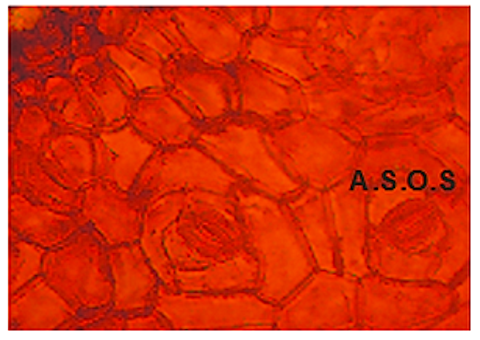
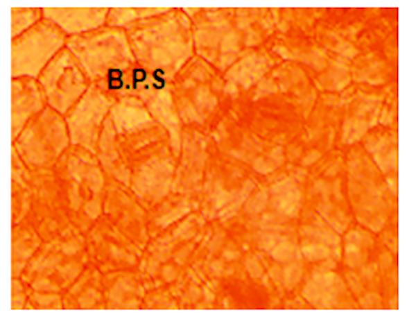
The anisocytic stomata was the commonest followed by brachyparacytic, anomocytic, staurocytic stomata and laterocytic stomatas respectively.
H. acetosella are distinguished on other species by having laterocytic stomata on both surfaces of leaves and parallel contiguous stomata are found on abaxial surface while in H. rosa-sinensis laterocytic is found only on adaxial surface.
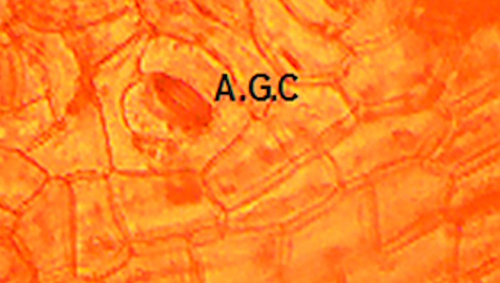
There are five different types of abnormal stomata, unopened stomatal pore, two stomata sharing one subsidiary cell, parallel contiguous stomata and aborted guard cell found in all the surfaces of the leaves and flowers.
In addition parallel contiguous stomata are found on adaxial surface of H. rosa-sinensis and abaxial surface of H. arnottianus flower. H. rosa-sinensis had five-armed trichome on the abaxial surface that helps in distinguishing it from other species studied.
The shape of epidermal cells, anticlinal cell walls, guard cell areas, stomatal index and trichomes varied. The results obtained could be used as diagnostic tool for plant identification and preparation of monograph on the species.

You must be logged in to post a comment.