Stomata Differentiation

Giving Voice to Stomata Differentiation

Torii K. U. (2007)

Cell 128(3): 419 – https://doi.org/10.1016/j.cell.2007.01.025

https://www.cell.com/cell/fulltext/S0092-8674(07)00123-7?_returnURL=https%3A%2F%2Flinkinghub.elsevier.com%2Fretrieve%2Fpii%2FS0092867407001237%3Fshowall%3Dtrue

The generation of asymmetry is a key event in many developmental processes, including cell differentiation, embryonic axis specification, and organogenesis. Recent research highlights the identification of factors needed to establish stomata during plant development and the role that timing plays in directing left-right asymmetry during nervous system development in zebrafish. Other recent studies elucidate unexpected connections between two kinds of asymmetry, cellular and organismal, and how asymmetry may be related to the regulation of chromatid segregation and DNA repair.

Giving Voice to Stomata Differentiation

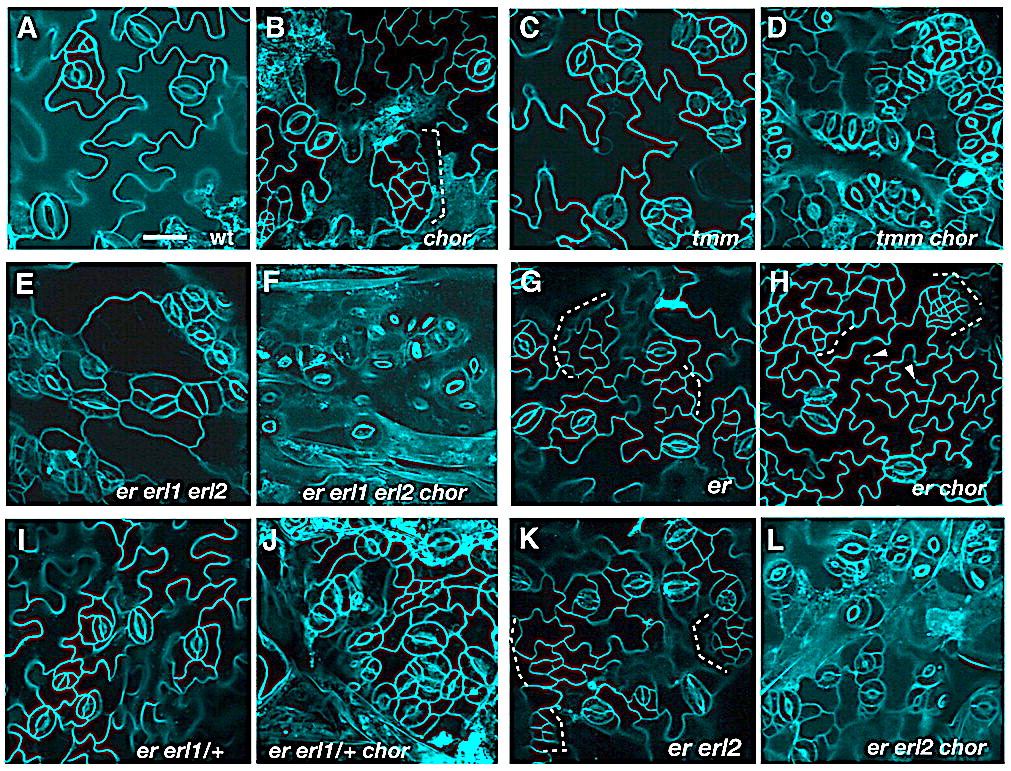
Stomata are the pores that mediate gas and water exchange in plants. They are formed by a symmetrical pair of guard cells that regulate the opening and closing of the pore, yet the specification of the guard cell precursor relies upon a series of asymmetric cell divisions beginning in a single meristemoid mother cell. Two groups (MacAlister et al., 2006; Pillitteri et al., 2006) now report that three related basic helix-loop-helix transcription factors (SPEECHLESS, MUTE, and FAMA) act sequentially in the lineage that gives rise to guard cells in Arabidopsis thaliana. As their names imply, loss of either SPEECHLESS or MUTE leads to seedlings that develop leaves devoid of stomata. In the first step of guard cell specification, SPEECHLESS facilitates the first asymmetric division of the meristemoid mother cell to establish the meristemoid. MUTE then terminates the subsequent rounds of asymmetric cell division to promote the differentiation of the meristemoid into a guard mother cell. The third factor FAMA, identified in a previous study, promotes the final transition from guard mother cell to guard cells. Future work may identify the targets regulated by these transcription factors. Of particular interest would be uncovering the genes regulated by SPEECHLESS that drive the asymmetric division of the meristemoid mother cell. The findings of MacAlister et al. and Pillitteri et al. also reveal a striking parallel between the differentiation of stomata and developmental processes in animals, including myogenesis and neurogenesis, which also require the sequential activity of related basic helix-loop-helix transcription factors.

C.A. MacAlister et al. (2006). Nature. Published online December 20, 2006. 10.1038/nature05491.

L.J. Pillitteri et al. (2006). Nature. Published online December 20, 2006. 10.1038/nature05467.

The similarities and uniqueness of two HD-ZIP IV genes in the specification of protodermal identity and stomatal differentiation beyond predetermined tissue layers

HDG2 is highly enriched in meristemoid population of stomatal cell lineages. (AHDG2 absolute and relative expression levels among wild-type and stomatal mutants enriched in specific epidermal cell populations. Absolute expressions (dark gray) are from ATH1 microarray data; relative expressions (light gray) are from qRT-PCR analysis. Data are mean values of triplicates; error bars indicate s.e.m. Col, wt; spch, pavement-cell only; mute scrm-D, overwhelmingly enriched in meristemoids; scrm-D, stomata-only epidermis. Below each graph are confocal images of cotyledons from corresponding genotypes. (B) Stomatal-lineage accumulation of HDG2 transcriptional reporter (HDG2pro::nls-3xGFP) in seedling epidermis. (C) Stomatal-lineage accumulation of HDG2 translational reporter (HDG2pro::HDG2-GFP) in 10-day-old abaxial cotyledon epidermis. Scale bars: 20 μm. (D) Expression levels of AtML1 and PDF2 compared with HDG2 among wild-type and stomatal mutants.

Arabidopsis homeodomain-leucine zipper IV proteins promote stomatal development and ectopically induce stomata beyond the epidermis

by Peterson K. M., Shyu C., Burr C. A., Horst R. J., Kanaoka M. M., Omae M., Sato Y., Torii K. U. (2013)

Department of Biology, University of Washington, Seattle, WA 98195, USA.

In Development. 140(9): 1924-1935 – doi: 10.1242/dev.090209 – Epub 2013 Mar 20 –

https://www.ncbi.nlm.nih.gov/pubmed/23515473

Abstract

The shoot epidermis of land plants serves as a crucial interface between plants and the atmosphere: pavement cells protect plants from desiccation and other environmental stresses, while stomata facilitate gas exchange and transpiration. Advances have been made in our understanding of stomatal patterning and differentiation, and a set of ‘master regulatory’ transcription factors of stomatal development have been identified. However, they are limited to specifying stomatal differentiation within the epidermis. Here, we report the identification of an Arabidopsis homeodomain-leucine zipper IV (HD-ZIP IV) protein, HOMEODOMAIN GLABROUS2 (HDG2), as a key epidermal component promoting stomatal differentiation. HDG2 is highly enriched in meristemoids, which are transient-amplifying populations of stomatal-cell lineages. Ectopic expression of HDG2 confers differentiation of stomata in internal mesophyll tissues and occasional multiple epidermal layers. Conversely, a loss-of-function hdg2 mutation delays stomatal differentiation and, rarely but consistently, results in aberrant stomata. A closely related HD-ZIP IV gene, Arabidopsis thaliana MERISTEM LAYER1 (AtML1), shares overlapping function with HDG2: AtML1 overexpression also triggers ectopic stomatal differentiation in the mesophyll layer and atml1 mutation enhances the stomatal differentiation defects of hdg2. Consistently, HDG2 and AtML1 bind the same DNA elements, and activate transcription in yeast. Furthermore, HDG2 transactivates expression of genes that regulate stomatal development in planta. Our study highlights the similarities and uniqueness of these two HD-ZIP IV genes in the specification of protodermal identity and stomatal differentiation beyond predetermined tissue layers.

Ligand-receptor pairs in stomatal development and beyond

Mix-and-match: ligand-receptor pairs in stomatal development and beyond

by Torii K. U. (2012)

Department of Biology, University of Washington, Seattle, WA 98195, USA.

===

In Trends Plant Sci. 17(12): 711-719 – doi: 10.1016/j.tplants.2012.06.013 – Epub 2012 Jul 21 –

https://www.ncbi.nlm.nih.gov/pubmed/22819466

Abstract

Stomata are small valves on the plant epidermis balancing gas exchange and water loss. Stomata are formed according to positional cues. In Arabidopsis, two EPIDERMAL PATTERNING FACTOR (EPF) peptides, EPF1 and EPF2, are secreted from stomatal precursors enforcing proper stomatal patterning. Here, I review recent studies revealing the ligand-receptor pairs and revising the previously predicted relations between receptors specifying stomatal patterning: ERECTA-family and TOO MANY MOUTHS (TMM). Furthermore, EPF-LIKE9 (EPFL9/Stomagen) promotes stomatal differentiation from internal tissues. Two EPFL peptides specify inflorescence architecture, a process beyond stomatal development, as ligands for ERECTA. Thus, broadly expressed receptor kinases may regulate multiple developmental processes through perceiving different peptide ligands, each with a specialized expression pattern. TMM in the epidermis may fine-tune multiple EPF/EPFL signals to prevent signal interference.

To understand the molecular mechanisms involved in cell-cell communication during stomatal development

Take a deep breath: peptide signalling in stomatal patterning and differentiation

by Richardson L. G., Torii K. U. (2013)

Department of Biology, University of Washington, Seattle, WA 98195, USA.

===

In J Exp Bot. 64(17): 5243-5251 – doi: 10.1093/jxb/ert246 – Epub 2013 Aug 30 –

https://www.ncbi.nlm.nih.gov/pubmed/23997204

Abstract

Stomata are pores in the leaf surface that open and close to regulate gas exchange and minimize water loss. In Arabidopsis, a pair of guard cells surrounds each stoma and they are derived from precursors distributed in an organized pattern on the epidermis. Stomatal differentiation follows a well-defined developmental programme, regulated by stomatal lineage-specific basic helix-loop-helix transcription factors, and stomata are consistently separated by at least one epidermal cell (referred to as the ‘one-cell-spacing rule’) to allow for proper opening and closure of the stomatal aperture. Peptide signalling is involved in regulating stomatal differentiation and in enforcing the one-cell-spacing rule. The cysteine-rich peptides EPIDERMAL PATTERNING FACTOR 1 (EPF1) and EPF2 negatively regulate stomatal differentiation in cells adjacent to stomatal precursors, while STOMAGEN/EPFL9 is expressed in the mesophyll of developing leaves and positively regulates stomatal development. These peptides work co-ordinately with the ERECTA family of leucine-rich repeat (LRR) receptor-like kinases and the LRR receptor-like protein TOO MANY MOUTHS. Recently, specific ligand-receptor pairs were identified that function at two different stages of stomatal development to restrict entry into the stomatal lineage, and later to orient precursor division away from existing stomata. These studies have provided the groundwork to begin to understand the molecular mechanisms involved in cell-cell communication during stomatal development.

SCREAM/ICE1 and SCREAM2 and stomatal differentiation


SCREAM/ICE1 and SCREAM2 specify three cell-state transitional steps leading to Arabidopsis stomatal differentiation.

by Kanaoka M. M., Pillitteri L. J., Fujii H., Yoshida Y., Bogenschutz N. L., Takabayashi J., Zhu J-K., Torii K. U. (2008)

  1. Masahiro M. Kanaoka
  2. Lynn Jo Pillitteri
  3. Hiroaki Fujii
  4. Yuki Yoshida
  5. Naomi L. Bogenschutz
  6. Junji Takabayashi
  7. Jian-Kang Zhu
  8. Keiko U. Torii

in Plant Cell 20:1775–1785. – 10.1105/tpc.108.060848. –

PubMed CentralView ArticlePubMed – Abstract/FREE Full Text

Abstract

Differentiation of specialized cell types in multicellular organisms requires orchestrated actions of cell fate determinants. Stomata, valves on the plant epidermis, are formed through a series of differentiation events mediated by three closely related basic-helix-loop-helix proteins: SPEECHLESS (SPCH), MUTE, and FAMA. However, it is not known what mechanism coordinates their actions.

Here, we identify two paralogous proteins, SCREAM (SCRM) and SCRM2, which directly interact with and specify the sequential actions of SPCH, MUTE, and FAMA. The gain-of-function mutation in SCRM exhibited constitutive stomatal differentiation in the epidermis.

Conversely, successive loss of SCRM and SCRM2 recapitulated the phenotypes of famamute, and spch, indicating that SCRM and SCRM2 together determined successive initiation, proliferation, and terminal differentiation of stomatal cell lineages.

Our findings identify the core regulatory units of stomatal differentiation and suggest a model strikingly similar to cell-type differentiation in animals.

Surprisingly, map-based cloning revealed that SCRM is INDUCER OF CBF EXPRESSION1, a master regulator of freezing tolerance, thus implicating a potential link between the transcriptional regulation of environmental adaptation and development in plants.

Antagonistic peptides and stomatal patterning


Competitive binding of antagonistic peptides fine-tunes stomatal patterning

by Lee J. S., Hnilova M., Maes M.Lin Y.-C. L.Putarjunan A.Han S.-K.Avila J.,Torii K. U. (2015)

in Nature 522,439–443(25 June 2015) doi:10.1038/nature14561

nature14561-f2
Figure 2: STOMAGEN overexpression on stomatal development in tmm hypocotyl epidermis with combinatorial loss-of-function in ER-family genes. – a–h, Representative confocal microscopy images of hypocotyl epidermis from 10-day-old light-grown transgenic Est::STOMAGEN (oestradiol-induced STOMAGEN) seedlings of tmm (a, b); tmm er (c, d); tmm erl1 erl2 (e, f); and tmm er erl1 erl2… – http://www.nature.com/nature/journal/v522/n7557/carousel/nature14561-f2.jpg

Abstract

During development, cells interpret complex and often conflicting signals to make optimal decisions. Plant stomata, the cellular interface between a plant and the atmosphere, develop according to positional cues, which include a family of secreted peptides called epidermal patterning factors (EPFs). How these signalling peptides orchestrate pattern formation at a molecular level remains unclear.

Here we report in Arabidopsis that Stomagen (also called EPF-LIKE9) peptide, which promotes stomatal development, requires ERECTA (ER)-family receptor kinases and interferes with the inhibition of stomatal development by the EPIDERMAL PATTERNING FACTOR 2 (EPF2)–ER module.

Both EPF2 and Stomagen directly bind to ER and its co-receptor TOO MANY MOUTHS. Stomagen peptide competitively replaced EPF2 binding to ER. Furthermore, application of EPF2, but not Stomagen, elicited rapid phosphorylation of downstream signalling components in vivo.

Our findings demonstrate how a plant receptor agonist and antagonist define inhibitory and inductive cues to fine-tune tissue patterning on the plant epidermis.

EPF1 and stomatal patterning

The secretory peptide gene EPF1 enforces the stomatal one-cell-spacing rule.

by Hara K., Kajita R.,

Torii K. U.,Torii

Bergmann D. C.,imgres

Kakimoto T.staff_image_1_2

(2007)

in Gene Dev 21:1720–1725 – – doi:10.1101/gad.1550707

http://genesdev.cshlp.org/content/21/14/1720.full

Abstract/FREE Full Text 

Abstract

Stomata are innovations of land plants that allow regulated gas exchange. Stomatal precursor cells are produced by asymmetric cell division, and once formed, signal their neighbors to inhibit the formation of stomatal precursors in direct contact.

We report a gene of Arabidopsis thaliana, EPIDERMAL PATTERNING FACTOR 1 (EPF1) that encodes a small secretory peptide expressed in stomatal cells and precursors and that controls stomatal patterning through regulation of asymmetric cell division.

EPF1 activity is dependent on the TOO MANY MOUTHS receptor-like protein and ERECTA family receptor kinases, suggesting that EPF1 may provide a positional cue interpreted by these receptors.

Cell-to-cell connectivity and stomatal patterning

F2.medium

CHOR is required to restrict stomatal cell-lineage divisions. (A-C) Promoter activity of TMM(proTMM::GUS-GFP) in 6-day-old cotyledon epidermis of wild type (A) and chor (B,C). In chor,clusters of small cells (dots) show high GFP signals. Pavement cells in chor occasionally show incomplete cytokinesis (arrowhead). (D-F) Expression of MUTE (proMUTE::MUTE-GFP) in 6-day-old cotyledon epidermis of wild type (D) and chor (E, F). Clusters of small stomatal-lineage cells are in brackets. MUTE expression in adjacent meristemoids (asterisks) predicts the eventual formation of clustered stomata. (G-I) Expression of SPCH (proSPCH::SPCH-GFP) in 3-day-old cotyledon epidermis of wild type (G) and chor (H, I). More cells express GFP signals in chor than in wild type. (J-L) Effects of spch on the excessive stomatal-lineage divisions in chor. Shown is 10-day-old cotyledon epidermis of spch (J) and spch chor (K,L). Small, highly divided cells are no longer produced in spch chor (K,L), whereas incomplete cytokinesis of pavement cells is still evident (K,L, arrowheads). Cell peripheries were highlighted by propidium iodide (PI; A-F,J-L) or FM4-64 (G-I). A-F, G-I, and J-L were taken under the same magnification. Scale bars: 20 μm.

Dysregulation of cell-to-cell connectivity and stomatal patterning by loss-of-function mutation in Arabidopsis CHORUS (GLUCAN SYNTHASELIKE 8).

by Guseman J. M., Lee J. S., Bogenschutz N. L., Peterson K. M., Virata R. E., Xie B., Kanaoka M. M., Hong Z., Torii K. U. (2010)

in Development 137:1731–1741. – doi: 10.1242/dev.049197 –

http://dev.biologists.org/content/137/10/1731 

F9.large
Fig. 9. Model of phenotypical consequences of the loss of CHOR (GSL8) and signaling receptors. (A) In wild type, stomatal cell-fate determinants (cyan) are contained in a precursor cell, and inhibitory signals (red T bar) prevent its neighbors from adopting a stomatal-lineage fate. (B) In chor, cell-fate determinants (cyan) may leak through plasmodesmata, which results in excessive stomatal entry divisions. The functional inhibitory signals, however, are able to prevent the stomatal differentiation of neighbors at some level. This results in modest stomatal clusters. Incomplete cytokinesis also occurs in chor. (C) In the absence of both CHOR (GSL8) and signaling receptors (such as TMM), the loss (or reduction at the threshold level in er erl1) of inhibitory signals together with the leakage of cell-fate determinants leads to catastrophic stomatal patterning defects. – http://d2qiws50qrj9uc.cloudfront.net/content/develop/137/10/1731/F9.medium.gif

Summary

Patterning of stomata, valves on the plant epidermis, requires the orchestrated actions of signaling components and cell-fate determinants.

To understand the regulation of stomatal patterning, we performed a genetic screen using a background that partially lacks stomatal signaling receptors.

F3.large

Genetic interactions of chor with stomatal patterning mutants. (A-L) Abaxial epidermis of rosette leaves (first pairs) from 12-day-old seedlings of the following genotypes: wild type (wt, A), chor (B), tmm (C), tmm chor (D), er erl1 erl2 (E), er erl1 erl2 chor (F), er (G), er chor (H), er erl1/+ (I), er erl1/+ chor (J), er erl2 (K) and er erl2 chor (L). chor in a wild-type background exhibits weak stomatal patterning defects and islands of small cells (dotted bracket; B), but the chormutation severely enhances stomatal clustering in tmm (C,D) and in the er erl1 erl2 triple mutant (E,F). er erl1/+ and er erl2 seedling leaves do not exhibit stomatal patterning defects (I,K); however, introduction of chor confers severe stomatal clustering defects (J,L). The er chordouble-mutant phenotype appeared additive, with increased small cells (dotted bracket) and incomplete cytokinesis (arrowheads, G,H). Images are taken under the same magnification. Scale bar: 20 μm.

Here, we report the isolation and characterization of chorus (chor), which confers excessive proliferation of stomatal-lineage cells mediated by SPEECHLESS (SPCH). chor breaks redundancy among three ERECTA family genes and strongly enhances stomatal patterning defects caused by loss-of-function in TOO MANY MOUTHS. chor seedlings also exhibit incomplete cytokinesis and growth defects, including disruptions in root tissue patterning and root hair cell morphogenesis.

CHOR encodes a putative callose synthase, GLUCAN SYNTHASE-LIKE 8 (GSL8), that is required for callose deposition at the cell plate, cell wall and plasmodesmata. Consistently, symplastic macromolecular diffusion between epidermal cells is significantly increased in chor, and proteins that do not normally move cell-to-cell, including a fluorescent protein-tagged SPCH, diffuse to neighboring cells.

Such a phenotype is not a general trait caused by cytokinesis defects.

Our findings suggest that the restriction of symplastic movement might be an essential step for the proper segregation of cell-fate determinants during stomatal development.

SPEECHLESS (SPCH), MUTE, and FAMA act during stomatal development.

Photo credit: NCBI

(A) Schematic diagram of stomatal development and the site of each stomatal bHLH gene action (arrows). Modified from Pillitteri et al.6 (B–E). The leaf epidermal phenotype of wild type

Stomatal Development

by Torii K. U., Kanaoka M. M., Pillitteri L. J., Bogenschutz N. L. (2007)

in Plant Signal Behav. –  v.2(4); Jul-Aug 2007 – PMC2634161 – 

http://www.ncbi.nlm.nih.gov/pmc/articles/PMC2634161/ 

Abstract

Stomata are microscopic pores on the plant epidermis that act as a major passage for the gas and water vapor exchange between a plant and the atmosphere. A pair of specialized guard cells work in concert to adjust pore size to maintain gas exchange while minimizing the water loss.

The formation of stomata requires a series of cell-fate transitions from an initial meristemoid mother cell (MMC), to a stem-cell-like precursor meristemoid, to a guard mother cell (GMC), and finally to terminally-differentiated guard cells.

Three closely-related Arabidopsis basic helix-loop-helix (bHLH) genes SPEECHLESS (SPCH), MUTE, and FAMA act sequentially at each key step to direct cell-fate transitions during stomatal development.

In this addendum, we propose that a three-step relay of the three bHLHs establishes the molecular framework for stomatal differentiation. Specific expression patterns as well as protein domain structure and dimerization partners of each stomatal bHLH protein may determine the specific function as a key switch in each regulatory node.