
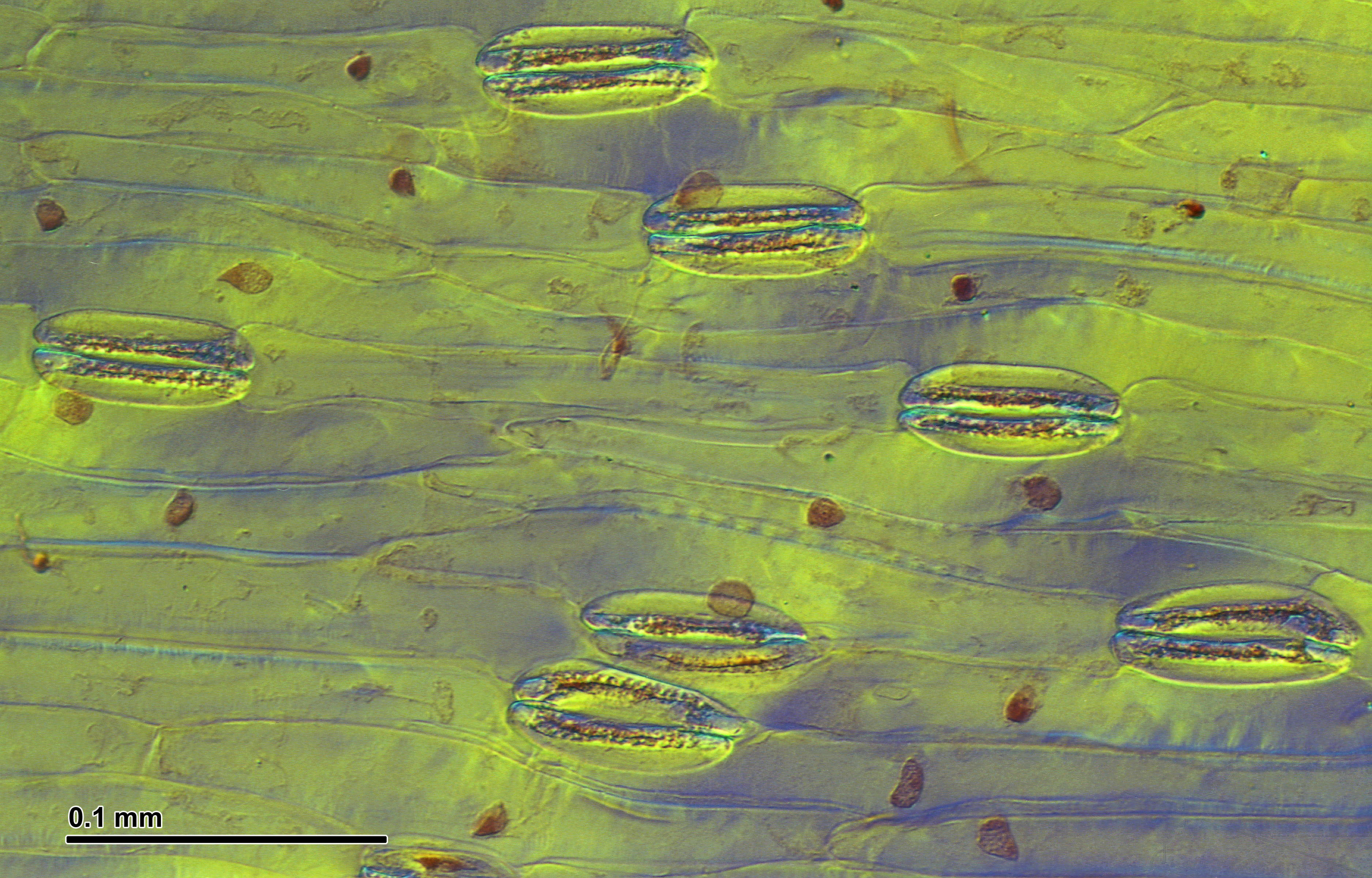
Optical microscopy technique:Differential interference contrast (Nomarski).
Magnification: 600x (for picture width 26 cm ~ A4 format).


Optical microscopy technique:Differential interference contrast (Nomarski).
Magnification: 600x (for picture width 26 cm ~ A4 format).


Bottom side of leaf with stomata
Taxonym: Carex melanostachya ss
Fischer et al. EfÖLS 2008 ISBN 978-3-85474-187-9
Location: Floridsdorf rail station, Vienna-Floridsdorf – ca. 160 m a.s.l.
Habitat: area below a pipe

by Lin C.-Y., Tan D.-Y. (2015)
Chen-Yi LIN, 1,2 AND Dun-Yan TAN, 1*
1 Key Laboratory of Western Arid Region Grassland Resources and Ecology, Ministry of Education, China; Xinjiang Key Laboratory of Grassland Resources and Ecology; College of Pratacultural and Environmental Science, Xinjiang Agricultural University, Ürümqi 830052, China
2 College of Forestry and Horticulture, Xinjiang Agricultural University, Ürümqi 830052, China
===
in Pak. J. Bot. 47(5): 1979-1988 –

Abstract
The genus Allium is comprised of more than 800 species, and although previous studies have been useful in identifying the species, there is a paucity of easy-to-observe morphological characters with which to distinguish them. Thus, we determined the micromorphological characteristics of the leaf epidermis of 43 species of Allium from Central Asia using light microscopy and evaluated their taxonomic significance.
Our study examined variability in epidermal cell shape and size and the stomatal apparatus.

The stomatal apparatus is ellipsoid, anomocytic and amphistomatic. The shape (rectangular or rhomboid) of epidermal cells, pattern (straight or arched) of anticlinal walls, and stomatal index are stable within a species, while there are differences among species that allow for species delimitation. Based on the shape and pattern of anticlinal walls of leaf epidermal cells, the 43 sampled species could be divided into three distinct types of epidermal cells: type 1, rhomboid cell shape and straight anticlinal walls; type 2, rhomboid cell shape and arched anticlinal walls; and type 3, rectangular cell shape and straight anticlinal walls.
These leaf epidermal micromorphological characters prove to be the taxonomic significance in distinguishing and delimitating species in Allium.

by Hofreiter A., Lyshede O. B. (2006)
Anton Hofreiter, Ole B. Lyshede,
===
in Botanical Journal of the Linnean Society 152(1): 73–90, https://doi.org/10.1111/j.1095-8339.2006.00540.x –
https://academic.oup.com/botlinnean/article/152/1/73/2420178
Abstract
The stomata appear only between the vascular bundles
by McKown K. H., Bergmann D. C. (2018)
1 Department of Genetics, Stanford School of Medicine, Stanford, CA 94305, USA.
2 Department of Biology, Stanford University, Stanford, CA 94305, USA; Howard Hughes Medical Institute, Stanford, CA 94305, USA.
===
in Curr Biol. 28(15): R814-R816 – doi: 10.1016/j.cub.2018.05.074 –
https://www.ncbi.nlm.nih.gov/pubmed/?term=30086309
Abstract
Stomata are adjustable valves through which gas and water exchange occur in plant leaves.
Here, McKown and Bergmann highlight the essential function and features of stomata from grasses.
by Wang W., Zhao N.-X. (2002)
| WANG Wei, ZHAO Nan-Xian, |
| South China Institute of Botany, The Chinese Academy of Sciences, Guangzhou 510650, China |
===
in Plant Sci J. 20: 343-349 –
http://www.whzwxyj.cn/EN/abstract/abstract1623.shtml
Abstract
The leaf epidermal structures of 27 species from 18 genera of Araceae and 1 species from Acoraceae were examined under light microscope,14 of which were observed with scanning electron microscope.
The stomatal apparatus types of Araceae vary from anomocytic, actinocytic to paracytic, cyclocytic and transitionary type between paracytic and cyclocytic; the numbers of subsidiary cells from zero to twelve.
Epidermal cells are nearly isodiametric in outline with straight, arched and undulate anticlinal walls.
Striate ornamentation occurs on periclinal walls of epidermal cells in some species. Although the stomatal apparatus types in Araceae are of little taxonomical significance at infra-family level of Araceae, the combined characters of stomatal apparatuses, the shape of anticlinal wall and ornamentation of cuticules in guard cells may be useful for species identification.
The separation of Acorus from Araceae is supported by leaf epidermal structures.

by Mantovani A., Pereira T. E. (2005)
André Mantovani, Instituto de Pesquisas Jardim Botânico do Rio de Janeiro, Programa Zona Costeira, Rua Pacheco Leão 915, CEP 22460-030, Jardim Botânico, Rio de Janeiro, Brasil,
Thaís Estefani Pereira, Bolsista Iniciação Científica PIBIC/CNPq.
===
in Rodriguésia 56 (88): 145-160 –
https://rodriguesia.jbrj.gov.br/FASCICULOS/rodrig56_88/10_Mantovani.pdf

ABSTRACT
Leaf and spathe anatomy of seven species and two varieties of the genus Anthurium (section Urospadix; subsection Flavescentiviridia) were analyzed. Plant material was collected from different locations in Brazil and cultivated under identical glasshouse conditions in the Rio de Janeiro Botanical Garden. Our attempt is to evaluate the diagnostic potential of leaf and spathe anatomy for taxonomic purposes. Leaves presented smooth cuticle, polygonal epidermal cells randomly disposed in paradermal view, periclinal divisions of epidermal cells in transversal view, non-raised stomata, collenchyma, sclerenchymatic bundle sheaths and raphides in the mesophyll. The spathe presented cuticularstriations; rectangular and elongated cellsin parallel rows; raised stomata; absence of collenchyma, raphides and sclerenchymatic bundle sheaths and presence of sclerenchyma as fibre caps under phloem. Clustering analysis based on leaf and spathe anatomical characters, revealed that the spathe can give a better resolution for segregation of species groups.
RESUMO
São apresentados dados relativos à anatomia da lâmina foliar e espata de sete espécies e duas variedades do gênero Anthurium pertencentes à seção Urospadix; subseção Flavescentiviridia. Os indivíduos foram coletados nos estados do Rio de Janeiro, São Paulo e Minas Gerais, e aclimatados no Instituto de Pesquisas Jardim Botânico do Rio de Janeiro. O objetivo deste estudo é comparar anatomicamente lâmina foliar e espata, visando detectar qual das duas estruturas é mais útil à diagnose taxonômica das espécies estudadas. Observa-se nas folhas a presença de cutícula lisa e células epidérmicas dispostas ao acaso, estômatos nivelados com a epiderme, divisões periclinais em células epidérmicas, além de ráfides no mesofilo e bainha esclerenquimática nos feixes vasculares. Já quanto à espata observa-se cutícula estriada, células alongadas e ordenadas de forma paralela, estômatos por vezes elevados, ausência de ráfides e presença de calota de fibras apenas junto ao floema, quando não ausentes. A análise de agrupamento para folha e espata revelou maior poder de resolução com base em caracteres anatômicos da espata; além dos grupos formados com base nos caracteres anatômicos da folha não serem consistentes taxonomicamente. Sugere-se portanto que a espata apresenta maior valor diagnóstico ao nível anatômico para subsidiar estudos taxonômicos do gênero Anthurium.

by Gomes Dias G. M., Rodrigues Soares J. D., Pasqual M., Lara Silva R. A., de Almeida Rodrigues L. C., Pereira F. J., de Castro E. M. (2014)
Gabrielen de Maria Gomes Dias1* , Joyce Dória Rodrigues Soares1 , Moacir Pasqual1 , Renata Alves Lara Silva1 , Luiz Carlos de Almeida Rodrigues2 , Fabricio José Pereira2Evaristo Mauro de Castro2
1 Federal University of Lavras (UFLA), Departamento of Agriculture, Plant Tissue Culture Laboratory, Postal Office 3037, 37200-000, Lavras- MG, Brazil
2 Federal University of Lavras (UFLA), Departamento of Biology, Postal Office 3037, 37200-000, Lavras- MG, Brazil
===
in Austral. J. Crop Science AJCS 8(8): 1160-1167 – ISSN:1835-2707 –
https://pdfs.semanticscholar.org/8a93/4a54f0acbe7f9c8e0dc43496a05d41d552ed.pdf
Abstract
The silicon can induce beneficial changes in plants, such as the further development of tissues and increased photosynthetic rate. Thus, studies on the anatomical changes resulting from in vitro culture are key to better understanding the development of micropropagated plants.
Therefore, this study was undertaken to evaluate the morphological and physiological differences in plants with the use of silicon added to the medium for the in vitro culture of Anthurium adreaenum cv. Rubi.
Nodal segments of seedlings were established in vitro and inoculated in Pierik medium supplemented with 30 g L-1 sucrose and solidified with 1.8 g L- 1 PhytagelTM. Different concentrations of sodium silicate (Na2SiO3 ) (0.0, 0.5, 1.0 and 2.0 mg L-1 ) were added to the medium. The experimental design was completely randomized with 30 repetitions. The segments were maintained for 100 days in a growth chamber under controlled conditions and evaluated anatomically and scanning electron microscopy (ultrastructurally) and for their photosynthetic capacities.
Medium containing 1.0 mg L-1 sodium silicate promoted the development of higher stomatal densities on the sheets. For the polar (31.38 µm) and equatorial (31.33 µm) diameter of the stomata of the abaxial leaf, the highest averages occurred in the treatment with 2.0 mg L-1 . Greater relative polar and equatorial diameters were estimated with a peak concentration of 1.2 mg L-1 . The increase in the sodium silicate concentration led to thinning of the abaxial and adaxial epidermis.
The thickness of the central rib had a sharp decrease up to 1.3 mg L-1 . For the mesophyll, the control displayed a higher thickness, whereas the addition of sodium silicate to the culture medium promoted a decrease. Seedlings grown in sodium silicate displayed significant differences, with increased photosynthetic and transpiration rates, stomatal conductance and internal CO2 concentrations. As for the ratio between the internal and external concentrations of CO2 , no significant differences were observed. The addition of sodium silicate resulted in increased epicuticular wax deposition and the formation of structures reserved for depositing calcium. Therefore, under in vitro conditions, the addition of sodium silicate to the culture medium affected the photosynthesis and leaf anatomy of A. andraeanum cv. Rubi, developing anatomical and physiological characteristics that contributed to the survival ex vitro.
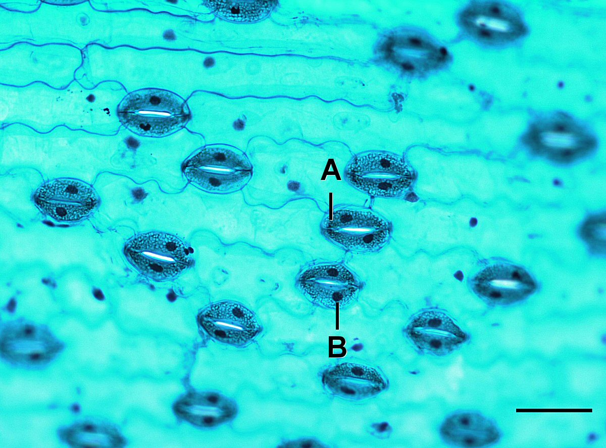
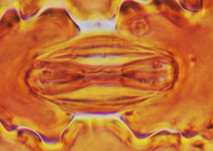
The leaves have stomata flanked on each side by two subsidiary cells parallel to larger stomatal axis (Fig 1A and B). Stomata showing two subsidiary cells displayed parallel to the larger stomatal axis are often classified as type brachyparacytic type (Castro et al., 2009). So, A. andraeanum stomata may be classified as paracytic type. Stomatal types in Araceae are very diverse and some species may show anomocytic, actinocytic, paracytic, and cyclocytic stomata (Wang and Zhao, 2002). Therefore, the brachypracytic stomata described by A. andraeanum may be an important characteristic for correct identification and was also reported by Mantovani et al. (2010) for some Anthurium species. Stomata guard cells showed a large number of chloroplasts and the usual bean-like shape (Fig. 1A). Despite the chloroplasts in stomatal guard cells is a very common anatomical characteristic (Castro et al., 2009) it was not highlighted by previous works in Anthurium anatomy (Wang and Zhao, 2002; Mantovani et al., 2010). Anticlinal cell walls are deeply sinuous in epidermal cells (Fig. 1A-D). Araceae leaves may show straight to very sinuous anticlinal cell walls in epidermal cells but a given species often show just one type (Keating, 2003). In nine Brazilian Anthurium species, Matovani et al. (2010) described from straight to undulated anticlinal cell walls. Therefore, the very sinuous anticlinal cell walls from A. andraeanum may be an important anatomical trait. The stomata are distributed only on the abaxial surface of leaves, classifying the se organs as hypostomatous (Fig 1). Hypostomatous leaves were also described in another Anthurium species by Mantovani et al. (2010) and Saito and Lima (2009).

You must be logged in to post a comment.