Stomata in Ericaceae

Photo credit: Google

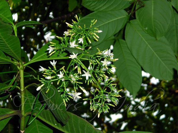
Cassiope_stellariana_15407.JPG

The taxonomic significance of certain anatomical variations on Ericaceae.THE ERICOIDEAE, CALLUNA AND CASSIOPE

by Watson L. (1964)

in J. Linn. Soc. (Bot.) 59: 111-125. – DOI: 10.1111/j.1469-8137.1964.tb07380.x – 

http://onlinelibrary.wiley.com/doi/10.1111/j.1469-8137.1964.tb07380.x/abstract

Summary

A review of the classical treatment of Ericaceae reveals how unconvincing are the usual definitions of the major groupings Ericoideae and Arbutoideae.

However, a survey of stomatal structure, stomatal distribution and pith structure supports the taxonomic soundness of the main body of the Ericoideae, which are further characterized by their distinctive habit. Calluna is exceptional among Ericoideae in pith structure and stomatal distribution, and differs from the others in various aspects of gross morphology.

There is a striking similarity between Calluna and Cassiope, a genus usually placed in the Arbutoideae; and there is little doubt that the two are closely related.

These conclusions lead to speculation about the origin and development of the Ericoideae in the minds of taxonomists, and about the mechanism by which closely similar genera become separated at the level of sub-family.

Giant stomata in Apocynaceae

Photo credit: Google

Tabernaemontana divaricata

Giant stomata in the family Apocynaceae

by Trivedi B. S., Upadhyay H. (1974)

in Current Science 43: 28, 29. –

https://eurekamag.com/research/000/102/000102832.php

batino
Alstonia macrophylla – http://www.flowersofindia.net/catalog/slides/Batino.jpg

Abstract

Giant stomata were observed in several species, including Alstonia macrophylla, Tabernaemontana divaricata and Strophanthus wightianus.

Conclusions on the significance of the leaf epidermis in Combretaceae


The significance of leaf epidermis in the taxonomy of the Combretaceae, Conclusions

by Stace C. A. (1980)

in Bot. J. Linn. Soc., (81) 327–339 – DOI: 10.1111/j.1095-8339.1980.tb01682.x – 

http://onlinelibrary.wiley.com/doi/10.1111/j.1095-8339.1980.tb01682.x/abstract

Abstract

The classification of the Combretaceae in relation to the anatomy of the leaf epidermis (especially to trichomes) is surveyed, with particular reference to Combretum. The new combination Calopyxis grandidieri (Drake) Capuron ex Stace is made and the new section Calopyxis sect. Petalantha Stace is described. A sectional review of Combretum is presented. The 69 sections are reduced to 45, and full bibliographic details, typification and synonymy are set out.


Stomata in Mimosa (dicots)

Scan 1Photo credit: Google

Mimosa pudica 

Epidermal structure and stomatal ontogeny in some Mimosaceae .

by Shah G. L., Parabia M. H., Kothari M. J.  (1972)

in Ann.Bot.,36:832-835. –

http://aob.oxfordjournals.org/content/36/4/823

Abstract

The structure of trichomes and stomata on leaflets of 21 species of the Mimosaceae are described.

Non-glandular trichomes in Mimosa pudica are of three types: unicellular, with a rounded thick-walled base and a terminal unicellular body, and multicellular. Capitate, clavate, or cylindric, 3–6-celled glandular hairs are observed on leaflets of Mimosa pudica only.

Leaflets are amphistomatic in all species except Adenanthera pavoninaCalliandra sp., Parkia biglandulosa, Pithecellobium dulce, and Samanea saman in which they are hypostomatic.

Only paracytic stomata are found in Leucaena leucocephala and Mimosa pudica. In the rest stomata are of more than one type. In spite of the diversity, the most frequent type in these species is paracytic. Anisocytic stomata, in all cases, are secondarily derived from paracytic ones by transverse or oblique wall formation in a subsidiary cell.

Similarly some stomata with one subsidiary cell are also secondary derivatives of the paracytic ones because of one of the subsidiary cells assuming the form of an epidermal cell.

The development has been traced in 14 species and that of paracytic stomata may be mesogenous or mesoperigenous, that of stomata with one subsidiary cell mesogenous but anomocytic stomata are ontogenetically perigenous. Occasionally a meristernoid is cut off from one of the subsidiary cells of a paracytic stoma. The organization of a stoma from such a meristemoid has been traced.

Scan

Scan 2

Scan 3

Scan 4

Stomata in Dioscorea (monocots)

Photo credit: Google

Dioscorea bulbifera

Scan

Some observations on the diversity of stomata and trichomes in six species of Dioscorea.

by Shah G. L., Gopal B. V. (1972)

Department of Botany, Sardar Patel University, Gujarat, India

in Ann. Bot. 36:997-1004. –

https://aob.oxfordjournals.org/content/36/5/997.short

Abstract

Stomata and trichomes are described on the leaves of six species of Dioscorea.

As many as six types were found in D. bulbifera and D. oppositifolia, four types in D.hispida and D. wallichii, three types in D. belophylla, but only two types in D. alata. Although there is a diversity of stomata even on the same surface, the predominant type is anomocytic in all the species except D. bulbifera in which it is tricytic. Rarely a stoma is also cyclocytic in D. bulbifera.

An increase in the number of subsidiary cells in paracytic, tricytic, or diacytic stomata takes place by the wall formation in them. Similarly a reduction in the number of subsidiary cells of a tetracytic stoma is the result of lateral subsidiary cells assuming the form of epidermal cells.

Abnormalities such as a stoma with one guard cell, degeneration of guard cells, and contiguous stomata are also met with.

The organization of different types of stomata is studied in D. bulbifera and D. wallichii and shown to be perigenous.

Capitate glandular hairs were seen on the leaves of D. belophylla, D. bulbifera, D. hispida, and D. wallichii but non-glandular, uniseriate, 3-celled trichomes were observed only in D. hispida.

Scan 1.jpeg

Scan 2

Stomata in Magnoliales

Photo credit: Google

Euptelea pleiosperma

Cuticular studies of Magnoliales

by Rao H. S. (1939) –  – Proc. Indian Acad. Sci. B 9: 99-116. – doi:10.1007/BF03050541 –

http://link.springer.com/article/10.1007/BF03050541

Summary

In the present work the question of the position of the Magnoliales from the point of view of their epidermal features has been taken up. This is rather a new line of inquiry. Florin’s criteria,viz., the haplocheile and syndetocheile types of stomatal development have been made use of.

According to this test the Magnoliales are seen to comprise a few synthetic forms, some possessing certain Cycadean features, the great majority the Bennettitalean, and a few the haplocheile gymnosperm features.

1280px-cercidiphyllum_japonicum2
https://upload.wikimedia.org/wikipedia/commons/thumb/3/31/Cercidiphyllum_japonicum2.jpg/220px-Cercidiphyllum_japonicum2.jpg

The aberrant epidermal features of Euptelea and Cercidiphyllum seem to be correlated with other primitive features of Trochodendraceæ already known.

The view has again been expressed that the progenitors of the angiosperms have to be sought for in that plexus of gymnosperms which gave rise to the Bennettitales, the syndetocheile Gnetales, the haplocheile Magnoliales and the majority of the Magnoliales (i.e., syndetocheile) along different lines of descent, the last being probably nearest related to the Bennettitales. From the point of view of epidermal studies, the Magnoliales do not represent a cosanguinary group, but indicate a convergence.

Stomata in Polemoniales (dicots)


Structure Ontogeny of Stomata in some Polemoniales.

by Patel R. C., Inamdar J. A. (1971)

in Annals of Botany: Vol. 35, 389-409. –

http://aob.oxfordjournals.org/content/35/2/389.abstract

Abstract

The ontogeny and structure of stomata in 22 genera and 51 species of the Polemoniales are described.

Five main types of stomata noticed are: anisocytic, anomocytic, diacytic, paracytic, and stomata with a single subsidiary cell.

Three modes of stomatal development: syndetocheilic or mesogenous, haplocheilic or perigenous, and meso-perigenous or syndeto-haplocheilic are observed.

Abnormalities seen are: stomata with single guard cells, arrested development and contiguous stomata variously oriented. Contiguous stomata result from adjacently placed meristemoids or readjustment during maturation. Stomata with a single guard cell are formed as a result of degeneration of one of the guard cells before or after pore formation.

The stomatal apparatus varies in different organs of a plant in form, number, orientation and arrangement of the subsidiary and also the surrounding cells.

Three lines leading to Polemoniales, Boraginales, and Solanales are distinct.

Scan

Scan 1

Stomata in Ficus (Moraceae)

Photo credit: Researchgate

Figure 6: Adaxial and abaxial leaf epidermis respectively in some members of subgenus Urostigma. a, b F. lutea (section Galoglychia, subsection Galoglychia); c, d F. sagittifolia (section Galoglychia, subsection Cyathistipulae); e, f F. elasticoides (section Galoglychia, subsection Crassicostae; g, h F. ingens (section Urostigma)

Leaf epidermal studies in some Nigerian species of Ficus L. (Moraceae).

by Ogunkunle A.T.J., Oladele, F.A (2008)

Ladoke Akintola University of TechnologyNigeria

Tunde Joseph OGUNKUNLE

tunde_ogunkunle

Tunde_OGUNKUNLE.png

University of Ilorin, Nigeria

Felix Oladele

felix_oladele
Felix_Oladele.jpg

in Plant systematics and Evolution, 214:209-221. – doi:10.1007/s00606-008-0044-9 –

http://link.springer.com/article/10.1007/s00606-008-0044-9

Abstract

Forty-two leaf epidermal features in 20 Nigerian species of Ficus, representing three of the four subgenera and four of the seven sections of the genus found in Africa have been examined.

Discontinuities in these characters clearly separated the three subgenera studied, namely, FicusSycomorus and Urostigma, and supported the earlier sectional classification of the subgenus Urostigma (i.e. sections Sycidium, Galoglychia and Urostigma).

The subsectional boundaries among the members of the section Galoglychia studied, however, had little backing from their leaf epidermal characteristics due to extensive overlap of the features.

The results of the present studies support the genetic basis of the qualitative and quantitative variations in the leaf epidermal cells, stomata and trichomes in the Nigerian Ficus and the present and potential applications of these features in diagnostic and taxonomic investigations in the genus are discussed.

Stomata in Senna (Caesalpiniaceae)


Foliar epidermal morphology of some Nigerian species of Senna (Caesalpiniaceae).

by Ogundipe O. T., Kadiri A. B., Adekanmbi O. H. (2009)

  • Department of Botany and Microbiology, University of Lagos, Lagos, Nigeria
12033132_626069234201429_2143130194094217812_n
Oluwatoyin Temitayo Ogundipe
Akeem_Kadiri2
Akeem Babalola Kadiri
olusola_adekanmbi
Olusola Helen Adekanmbi

in Indian J. Sci. & Tech. 2(10): 5-9. –

http://www.indjst.org/index.php/indjst/article/view/30709

Abstract

A comparative foliar epidermal and petiole anatomical morphology of six commonly used pharmacodynamic species of Senna in Nigeria was investigated with a view to elucidating their taxonomic significance and present complementary data which would aid the recognition of the species.
The cell shape may be polygonal and irregular on both surfaces and either polygonal or irregular on any of the two surfaces of the leaf. Anticlinal wall is either straight or undulate and curved. Usually, the epidermal cell size is more on the adaxial surface and the leaf is either amphistomatic or hypostomatic.
Trichomes are usually slender acicular to conical and glandular or non-glandular. The vascular system of the petiole is arranged in a spaced arc.
Based on this an indented dichotomous key is presented to distinguish the species.

Stomata in Acalypha (dicots)

Photo credit: Google

Acalypha segetalis

Leaf Epidermis Morphology of West African species of the genus Acalypha (Euphorbiaceae).

by Kadiri A. B., Olowokudejo J. D., Ogundipe O. T. (2009)

University of Lagos, Nigeria

Akeem Babalola Kadiri,

Akeem_Kadiri2
Akeem Babalola Kadiri

James Dele Olowokudejo,

james_olowokudejo
James Dele Olowokudejo

Oluwatoyin Temitayo Ogundipe

12033132_626069234201429_2143130194094217812_n
Oluwatoyin Temitayo Ogundipe

in Botanica Lithuanica. 15(2): 65-78. –

http://www.botanika.lt/botli_en.htm

http://connection.ebscohost.com/c/articles/47556556/leaf-epidermis-morphology-west-african-species-genus-acalypha-euphorbiaceae

Abstract

Comparative foliar epidermal characteristics of 15 species of Acalypha L. in West Africa are analysed using both light and scanning electron microscopy.

Presence of hypostomatic leaves supports the series accepted by Bentham and Hooker (ser. Pantogynae-Acrogynae and ser. Pantogynae- Pleurogynae) except A. wilkesiana. In taxa of other series amphistomatic leaves were recorded.

Among other taxonomically useful features of the genus are polygonal and irregular epidermal cell shapes, curved and undulate anticlinal walls, presence of regular to irregular striae and deposition of waxy flakes on the leaf surfaces.

A. ceraceopunctata and A. segetalis can be distinguished from other species by the presence of peltate scale and only paracytic stomata respectively.

An artificial bracketed dichotomous key is presented to delimit species of the genus Acalypha.