
Photo credit: Google
Caesalpinia spinosa
Pericarpial diversity in Caesalpinia pods used as tanning material
Stant M. V. (1972)
Margaret V. Stant
in Bot. J. Linn. Soc.65: 313-334


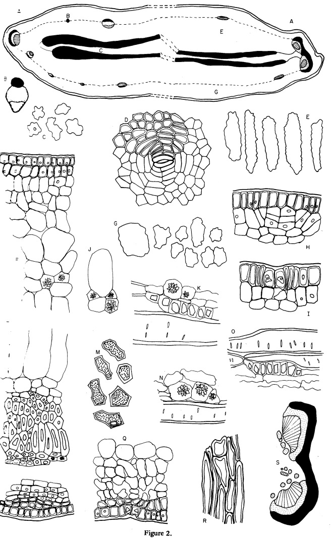


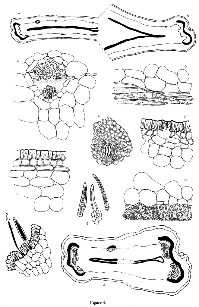

Photo credit: Google
Caesalpinia spinosa
Stant M. V. (1972)
Margaret V. Stant
in Bot. J. Linn. Soc.65: 313-334
Photo credit: Google
Ouratea spectabilis
by Arens T. (1968)
Cadeira de Botânica da Faculdade de Fil.Est. de São PauloBrasil
in Protoplasma (Wien) 66: 403-411 –
https://link.springer.com/article/10.1007/BF01255867
Zusammenfassung
An den lebenden unbehandelten Spaltöffnungen von Ouratea spectabilis wurde eine vom Spalt ausgehende Radialstruktur beobachtet.
Dieselbe läßt sich mit verschiedenen Farbstoffen darstellen und ist nicht mit einer Cuticularstreifung identisch.
Es handelt sich um Ektodesmen, die infolge ihrer besonderen Eigenschaften (Größe usw.) ohne Anwendung spezieller Methoden sichtbar sind.
Mit der Methode von Bancher, Hölzl und Klima läßt sich zeigen, daß aus ihnen Flüssigkeitströpfchen abgeschieden werden können.
Mit dem Transpirationswasser aufgenommene Salze (Thallium und Mangan) reichern sich im Zellumen und in diesen Ektodesmen an.
Sie sind offenbar die Bahnen der peristomatären Transpiration.
—————————–
Radial structures in the stomata of Ouratea spectabilis (Mart.) Engl.
Summary
In living, untreated stomata of Ouratea spectabilis a radial structure originating in the stomatal opening was observed, which may be demonstrated by means of various stains.
It is not identical with a cuticular striation.
These structures are shown to be ectodesms visible without use of special methods because of their peculiar properties (size etc.).
The method of Bancher, Hölzl and Klima allows to demonstrate that these ectodesms may secret fluid droplets. Salts (thallium and manganese) taken up with the transpiration stream are concentrated in the cell lumen and in the ectodesms. Obviously, the ectodesms are the pathways of peristomatal transpiration.
Photo credit: Google
Nelsonia campestris

by Ahmad K. J. (1974)
in Bot. J. Linn. Soc. 68: 73-80 –
This paper represents a modified portion of a thesis accepted for the Ph.D. degree by the University of Lucknow.
Abstract
The foliar epidermis and cuticle of Staurogyne longifolia (Nees) Kuntze, Elytraria acaulis (L.f.) Lindau var. acaulis, E. acaulis var. lyrata (Nees) Bremek. and Nelsonia campestris R.Br, have been investigated, revealing broad similarities with those of the rest of the Acanthaceae;
the presence of diacytic stomata in the Nelsonioideae is evidence of its affinity with the Acanthaceae in general, while the presence of panduriform glandular hairs and the absence of the cystoliths in Nelsonioideae indicate its particular affinity with the Thunbergioideae.
Substantial evidence is provided to support the retention of Nelsonioideae as a subfamily of the Acanthaceae, rather than its transfer to the Scrophulariaceae.
Photo credit: Google
Sarcandra glabra
Ramji M. V. (1964)
University of Madras, India
in Proc. Indian Acad. Sci, Bangalore 59: 360-364

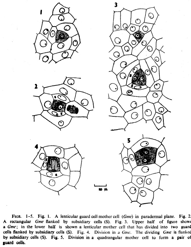
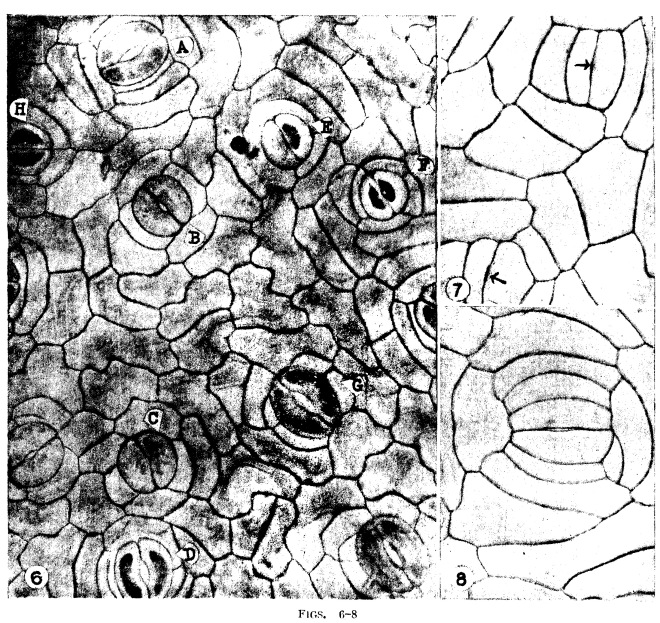
Photo credit: Google
Sesuvium portulacastrum
by Ramayya N., Rajagopal T. (1971)
Osmania University, Hyderabad, India
in J. Indian bot. Soc. 50: 355-362
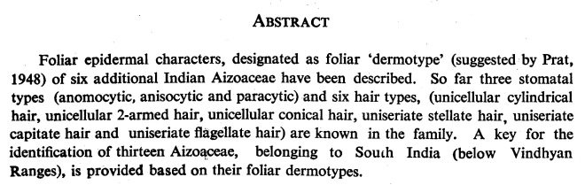

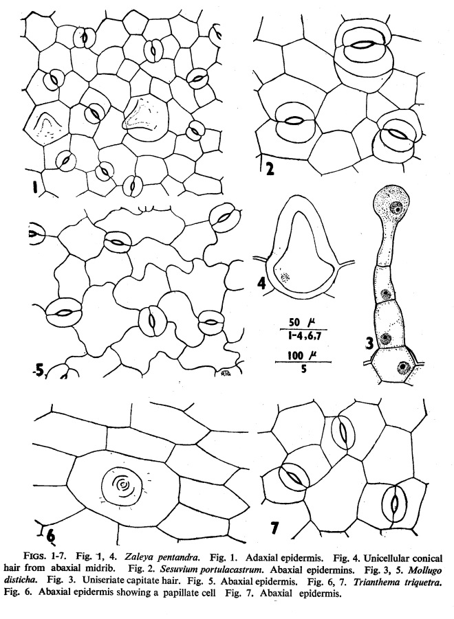
Photo credit: Google
Citrus limon
by Reed H. S., Hirano E. (1931)
H. S. Reed, University of California, USA
E. Hirano, Tottori Agric. College, Japan
in J. Agric. Res. 43(3): 209-222


par Rousteau A. (1981)
Université Pierre et Marie Curie, Paris, France
Alain Rousteau
in Rev. gén. Bot. 88: 23-31

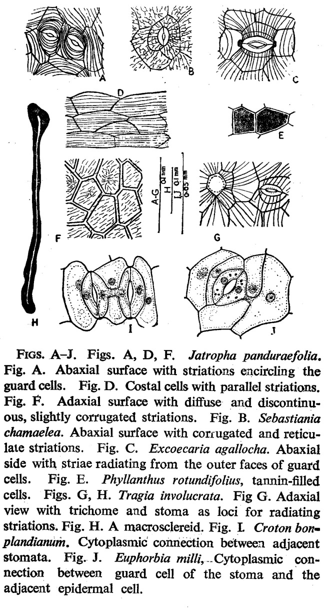
Photo credit : Google
Sebastiana chamaelea
by Rao P. N., Raju V. S. (1975)
Andhra University, Waltair, India
Piratla Narasimha Rao, V. Satyanarayana Raju
in Curr. Sci. 44: 750-752

by Rao P. N., Raju V. S. (1975)
Andhra University, Waltair, India
Piratla Narasimha Rao, V. Satyanarayana Raju
in Curr. Sci. 44: 594-596

Photo credit: Google
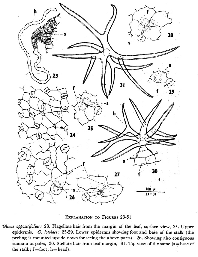
Glinus lotoides var. virens
by Ramayya N., Rajagopal T. (1968)
in Journ. Osmania University (Sci.) , Golden Jubilee Volume, Osmania University, Hyderabad. – pp. 147-160


You must be logged in to post a comment.