ABA, CO2 and Ca2+ in stomata


Guard cell ABA and CO2 signaling network updates and Ca2+ sensor priming hypothesis.

by Israelsson M., Siegel R. S.,

 Young J.,Jared_Young3

 Hashimoto-Sugimoto M., Iba K.

 Schroeder J. I.julian-schroeder-151015

(2006)

 in Current Opinion in Plant Biology 9654663. – doi:10.1016/j.pbi.2006.09.006

CrossRefMedlineWeb of Science

http://cel.webofknowledge.com/InboundService.do?product=CEL&SID=Q1KxcDMHKXP3N1gwe6c&UT=WOS%3A000242057800015&SrcApp=Highwire&action=retrieve&Init=Yes&SrcAuth=Highwire&Func=Frame&customersID=Highwire&IsProductCode=Yes&mode=FullRecord

Abstract

Stomatal pores in the epidermis of plants enable gas exchange between plants and the atmosphere, a process vital to plant life. Pairs of specialized guard cells surround and control stomatal apertures.

Stomatal closing is induced by abscisic acid (ABA) and elevated CO2 concentrations. Recent advances have been made in understanding ABA signaling and in characterizing CO2 transduction mechanisms and CO2 signaling mutants. In addition, models of Ca2+-dependent and Ca2+-independent signaling in guard cells have been developed and a new hypothesis has been formed in which physiological stimuli are proposed to prime Ca2+ sensors, thus enabling specificity in Ca2+-dependent signal transduction.

Stomata in Boraginaceae


Biosystematic Studies in Heliotropium indicum, Trichodesma indicum and T. zeylanicum of Boraginaceae

by Kumar A., Kumar B. (2016)

in Int.J.Curr.Microbiol.App.Sci (2016) 5(4): 720-729 – DOI: http://dx.doi.org/10.20546/ijcmas.2016.504.083 –

http://www.ijcmas.com/5-4-2016/Ashwini%20Kumar%20and%20Baidyanath%20Kumar.pdf 

Abstract

In the present observation biosystematics of three species of Boraginaceae viz., of Heliotropium indicum, Trichodesma indicum and Trichodesma zeylanicum was studied. All the three species showed a marked difference in leaf and inflorescence morphology, stomatal behaviour, epidermal trichome, pollen grain characteristics and stem anatomy, hypodromous and brochidodromous venation of their leaves.

Leaves were landeolate to ovate. The inflorescence colour was pale blue to white. Length of inflorescence was found to be 4.0 to 6.5. The stomatal Index in these three species varied greatly. T. indicum and T. zeylanicum showed anomocytic stomata with 16mm in length and 12mm width and Stomatal Index was 34.88 and 44.68 respectively.

Heliotropium indicum exhibited anisocytic stomata which were 12mm long and 10mm wide. The Stomatal Index was 32.65.

All the three species showed the presence of unicellular glandular hairs which differed in forms. All the three species showed tricolpate pollen grains, but differed in their form and size. All the three species showed more or less similar stem anatomical features with differences only in their hypodermal layers. Numerical analysis with reference to morphological, stomatal, pollen grains, trichomes, stem anatomy and stomatal index was carried out for 31 characters.

It was found that Heliotropium indicum showed similarity with Trichodesma indicum in only eight characters and with Trichodesma zeylanica in only nine charaters. Trichodesma indicum and T. zeylanicum showed resemblances in twenty two characters.

On the basis of matching coefficient it was observed that Heliotropium indicum showed only 25.80% resemblance with T. indicum, and 29.03% with T. zeylanicum.Trichodesma indicum and T. zeylanicum showed 70.96% resemblances, and therefore might be considered as same species of the genus Trichodesma.

Stomata in Bryophyllum


Epidermal structure and stomatal types in vegetative and reproductive organs of three species of Bryophyllum

by Jeong W.-G., Kim C. S. (1987)

in Korean J. Bot. 30. (1): 43-58. –

http://agris.fao.org/agris-search/search.do?recordID=KR19890035333

Abstract
The epidermal structure and stomatal types in vegetative and reproductive organs of three species of Bryophyllum (B. crenatum, B. diagremontian, B. tubiflorum) were described.
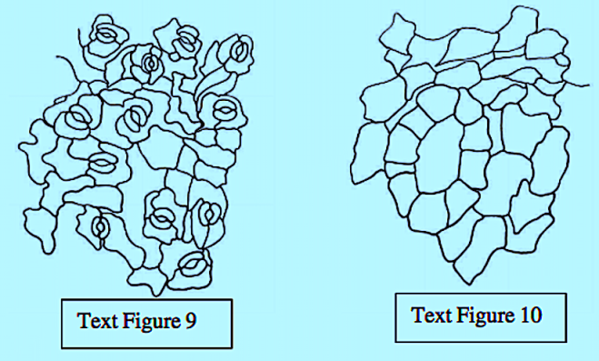
The epidermal cells were polygonal, isodiametric, and rectangular in the leaves and stems, styles, and ovaries. These cells were commonly thick, and arched or sinuous in the leaves, epiphylous buds, petals and ovaries. They were straight in the stems, petioles, pedicels, and peduncles. In both vegetative and reproductive organs, the subsidiary cell walls were commonly thin and mostly arched in all the organs.
The great majority of the mature stomata in all the organs were helicocytic type with a helix of four to six subsidiary cells. The mature stomata varied from organ to organ with regard to the number and arrangement of subsidiary cells.
The ontogenetic type of stomata in all the organs was mostly helico-eumesogenous type. This type was subdivided into three subtypes such as parahelico-eumesogenous, anomohelico-eumesogenous, and diahelico-eumesogenous stomata on the basis of the division angle of the guard mother cell.
Sometimes, the aniso-eumesogenous type was found in various organs. This type was subdivided into three subtypes such as paraniso-eumesogenous, anomoaniso-eumesogenous, and dianiso-eumesogenous stomata.
The tetra-eumesogenous and duplotetra-eumesogenous types were rarely found; the former in the leaf of B. crenatum and the latter in the leaf of B. diagremontiana.
An omomeristic patterns in the mesogenous category of stomatal types was observed in a few organs of all the materials.
A new stomatal type with tetra-eumesogenous stoma within a girdle of three subsidiary cells of aniso-eumesogenous in the leaf of B. diagremontiana was firstly observed in the vascular plants. This stoma was termed the cotetra-aniso-eumesogenous type.
Abnormal stomata such as aborted stomata, single guard cells, stoma with a constricted part in the middle of large guard cells, and arrested stomata were found in the various organs of all the materials.

Stomata in 4 monocots


Stomatal Pattern in Four Species of Monocotyledons

by Charlton W. A. (1988)

in Ann Bot (1988) 61 (5): 611-621. – 

http://aob.oxfordjournals.org/content/61/5/611 

Abstract

The distribution of stomata has been investigated in the leaf epidermis of Chlorophytum comosum, Galanthus nivalis, Schizostylis coccinea and Scilla lancifolia. The epidermis was considered to consist of units of construction of two kinds: type A, a long epidermal cell with a stoma at its distal end, and type B, a long epidermal cell without an associated stoma.

Except in Scilla, the probability of an epidermal unit being type A increases approximately with its length. Considering the epidermis as rows of units, alternating sequences of type A and type B do not occur randomly along the rows. In Chlorophytum, Galanthus and Schizostylis, both type A and B units tend to be aggregated into longer sequences than would be expected on a random basis.

It is suggested that homoeogenetic induction (i.e. of like by like) may be occurring during development. No case can be made for homoeogenetic induction of units in Scilla. There is a slight tendency to periodicity of distribution of type A units in GalanthusSchizostylis and Scilla, but this does not seem to represent a primary element of pattern.

There is interaction between rows in the sense that unit ends (transverse walls) tend to avoid those in neighbour rows; this affects the relative distribution of stomata, but there is no evidence of any direct interaction between stomata in different rows.

Types of stomata


What types of stomata are found in plants ?

by Anonymous (x)

in https://www.reference.com/science/types-stomata-found-plants-6199e4c98fa2cdba

What types of stomata are found in plants?

QUICK ANSWER

As of 1970, plant stomata have been divided into eight types: actinocytic, anisocytic, anomocytic, cyclocytic, diacytic, hexocytic, paracytic and tetracytic.

A typical stomata formation found in the dicotyledons, or flowering plants, is the anisocytic type that is comprised of a stoma surrounded by three cells of different sizes. The stomata are the microscopic pores on the epidermal layers of land plants that enable them to exchange oxygen, a by-product of photosynthesis, for carbon dioxide.

The presence of stomata on the external portions of land plants is a critical factor in their survival. The stomatal pores protect a plant by opening and closing in response to changes in environmental conditions. In times of drought, for example, the stomatal pores will close to keep water inside the plant and to prevent wilting and dehydration. At the same time, the stomata regulate the carbon dioxide and oxygen gas exchange portion of the respiratory process of land plants that plays a vital role in the global environment.

Read the full article: Reference


Stomata in Malvaceae


Stomatal studies on some selected plants of Malvaceae

by Chachad D. P., Vaidya M. (2016)

Screen Shot 2018-05-27 at 21.19.51

in World Journ. Pharmac. Research –  5 (3): 1060-1068 –

www.wjpr.net  article_wjpr_1456806564.pdf

https://www.academia.edu/23957454/STOMATAL_STUDIES_ON_SOME_SELECTED_PLANTS_OF_MALVACEAE

ABSTRACT

Malvaceae family is well known for its economic importance. Plants like Hibiscus esculantus is commonly used as a vegetable called ‘Okra’ Adensonia digitata, known as baobab tree has a lot of medicinal potential.

Bombax ceiba yields Kapok which is a very fluffy material made from the abundant silken hairs that are attached to the ripe seeds of several species in the silk cotton family. As mentioned in the literature, plants belonging to Malvaceae are economically important not only with respect to edible purposes but are also known to be exploited for their medicinal potential.

Anatomy is a very useful tool for identification (taxonomy). Epidermal characters can prove to be of importance with respect to identification of a particular plant species. This can also provide reliable information in standardisation of an ayurvedic/herbal drug.


Abutilon ranadei
: Upper epidermis shows anomotetracytic, cyclocytic type of stomata withkidney shaped guard cells. Epidermal cells are irregular with wavy margin. Text Figure 1Lower epidermis shows anomotetracytic, anisocytic type of stomata with kidney shapedguard cells. Epidermal cells are irregular with wavy margin. 
 Screen Shot 2018-05-27 at 21.24.46
Adansonia digitata
: Upper epidermis shows polygonal cells which form glands with wavy margin. Hypostomatic. Text Figure 3.Lower epidermis shows anisocytic, anomotetracytic and cyclocytic type of stomata withkidney shaped guard cells. Epidermal cells are irregular with wavy margin. Text Figure 4.
Screen Shot 2018-05-27 at 21.27.15
Bombax ceiba
: Upper epidermis shows tetra-pentagonal cells. Hypostomatic. Text Figure 5.Lower epidermis shows anisocytic, anomotetracytic and cyclocytic type of stomata with kidney shaped guard cells. Epidermal cells are irregular with wavy margin. Text Figure 6
Screen Shot 2018-05-27 at 21.30.18
Bombax malabaricum
: Upper epidermis shows pentagonal to hexagonal epidermal cellswhich are irregularly scattered. Hypostomatic. Text Figure 7.Lower epidermis shows anisocytic, & paracytic type of stomata with kidney shaped guardcells. Epidermal cells are irregular with wavy margin. Text Figure 8
Screen Shot 2018-05-27 at 21.32.40
Hibiscus decusseta
: Upper epidermis shows anomocytic, paracytic & anisocytic type ofstomata with kidney shaped guard cells. Epidermal cells are irregular with wavy margin. TextFigure 9.Lower epidermis shows pentagonal cells, cells arranged to form glands. Hyperstomatic. TextFigure 10
Screen Shot 2018-05-27 at 21.34.39

ABA and MeJA signaling in Arabidopsis stomata


Cytosolic alkalization and cytosolic calcium oscillation in Arabidopsis guard cells response to ABA and MeJA.

by Islam M. M., Hossain M. A., Jannat R., Munemasa S., Nakamura Y., Mori I. C., Murata Y. (2010)

in Plant Cell Physiol. 51: 1721–1730 – doi: 10.1093/pcp/pcq131.

[PubMed]

http://www.ncbi.nlm.nih.gov/pubmed/20739306http://www.ncbi.nlm.nih.gov/pubmed/20739306

Abstract

Abscisic acid (ABA)- and methyl jasmonate (MeJA)-induced stomatal closure are accompanied by cytosolic alkalization in guard cells. However, it remains to be clarified how the alkalization functions in not only ABA signaling but also MeJA.

We investigated cytosolic alkalization in guard cells during ABA-, MeJA- and Ca(2+)-induced stomatal closure of wild type, abi1-1, abi2-1, ost1-2 and coi1 using a pH-sensitive fluorescent dye, BCECF-AM.

ABA induced cytosolic alkalization in guard cells of wild-type and coi1 but not in ost1-2 guard cells whereas MeJA elicited cytosolic alkalization in wild-type and ost1-2 guard cells but not in coi1. Neither ABA nor MeJA induced cytosolic alkalization in abi1-1 and abi2-1 guard cells.

Exogenous Ca(2+) induced stomatal closure accompanied by cytosolic alkalization in guard cells of wild-type, abi1-1, abi2-1, ost1-2 and coi1 plants.

An agent to acidify cytosol, butyrate, suppressed Ca(2+)-induced cytosolic alkalization and ABA-, MeJA- and Ca(2+)-induced cytosolic Ca(2+) oscillation in wild-type guard cells to prevent stomatal closure.

These results indicate that cytosolic alkalization and cytosolic Ca(2+) oscillation coordinately function in ABA and MeJA signaling in Arabidopsis guard cells.

Stomatal movements, pH and Ca.


Changes in cytoplasmic pH and calcium in guard cells precede stomatal movements.

by Irving H. R., Gehring C. A., Parish R. W. (1992)

in Proc. Natl Acad. Sci. USA, 89, 17901794. – 

CrossRef |PubMed | [PMC free article]

Abstract

Stomatal opening is induced by indoleacetic acid (IAA), cytokinins, and fusicoccin (FC), whereas stomatal closure is induced by abscisic acid (ABA).

To test the effect of these growth regulators on guard cell cytosolic Ca2+ ([Ca2+]cyt) and pH (pHcyt), epidermal strips were taken from the lower side of leaves of the orchid Paphiopedilum tonsum and were loaded with acetomethoxy-esterified forms of the Ca2+ indicator fluo-3 or the pH indicator 2′,7′-bis(2-carboxyethyl)-5(6)carboxyfluorescein.

Basal [Ca2+]cyt ranged from 0.05 to 0.3 M and was 0.22 +/- 0.015 (n = 21). Increases in both [Ca2+]cyt and pHcyt were observed in guard cells after application of 10-100 M ABA to open stomata, and these preceded stomatal closure.

The increase in [Ca2+]cyt ranged from 1.5- to 3-fold and was seen in 7 of 10 experiments. Guard cell alkalinization began within 2 min of ABA treatment and continued for the next 8 min. The increase ranged from 0.04 to 0.3 pH unit and was seen in 13 of 14 experiments. Guard cell [Ca2+]cyt increased, whereas pHcyt decreased after treatment of closed stomata with IAA, kinetin, or FC.

In response to 50-100 M IAA, [Ca2+]cyt increased 1.5- to 2-fold in all cases, and pHcyt decreased 0.2-0.4 unit within 5 min in 7 experiments. Within 12 min, 10-100 M kinetin caused [Ca2+]cyt to increase in 28 of 34 experiments (1.3- to 2.5-fold) and pHcyt fell 0.1-0.4 unit in 15 of 17 treatments. The response to 10-50 M FC was similar in both time and magnitude.

These results show that stomatal opening is accompanied by an increase in [Ca2+]cyt and cytosolic acidification in the guard cells, whereas stomatal closure is preceded by an increase in [Ca2+]cyt and cytosolic alkalinization in the guard cells. The order of these events is still uncertain, but changes in pHcyt are correlated with stomatal movement, and these changes may be an important factor in the regulation of guard cell movement.

Phototropin-mediated signaling process of stomatal guard cells


Phototropin signaling and stomatal opening as a model case.

by Inoue S., Takemiya A., Shimazaki K. (2010)

in Curr. Opin. Plant Biol. 13, 587593 (2010). –  doi:10.1016/j.pbi.2010.09.002

CASPubMedArticle

Abstract

Phototropins are plant-specific light-activated receptor kinases that regulate diverse blue-light-induced responses, and serve to optimize plant growth under various light environments. Phototropins undergo autophosphorylation as an essential step for their signaling and induce a variety of tissue-specific or organ-specific responses, but the divergent mechanisms for these responses are unknown.

It is most likely that the phototropins generate a specific output after the event of autophosphorylation.

In this report, we will review the common steps of phototropin signaling and the numerous interactive proteins of phototropins, which may act as signal transducers for the diverse responses. We also describe the phototropin-mediated signaling process of stomatal guard cells and its crosstalk with abscisic acid signaling.

Ca leads to the suppression of stomatal opening.


Calcium inhibits ion-stimulated stomatal opening in epidermal strips of Commelina communis L.

by Inoue H., Katoh Y. (1987)

in Journal of Experimental Botany 38, 142149. –

CrossRefCAS |

http://jxb.oxfordjournals.org/content/38/1/142

Abstract

Ca2+ suppressed both the ion-stimulated stomatal opening and H+extrusion of pre-illuminated epidermal strips isolated from Commelina communis L.

In the absence of Ca2+, the rate of H+ release was 18 nmol H+ cm–2 h–1 per epidermal strip unit area in 150 mol m–3 KCL at pH 7·4. Half-maximum inhibition of stomatal opening was observed with 220 mmol m–3 of Ca2+.

The hexavalent dye, ruthenium red, showed concentration-dependent prevention of the inhibition by Ca2+ of the ion-stimulated stomatal opening. The effect of ruthenium red was non-competitive, and the K1 for the calcium inhibition was found to be 3·6 mmol m–3. The calcium inhibition of H+ extrusion was also prevented by ruthenium red.

These results suggest that Ca2+ inhibits the activity of electrogenic H+ translocating ATPase of the guard cell plasma membrane and leads to the suppression of stomatal opening.