
Guard cell ontogeny in leaf stomata of the fern Ophioglossum petiolatum
Peterson R. L., Hambleton S. (1978)
R. L. Peterson, Sarah Hambleton,
in Canadian Journal of Botany 56: 2836–2852 – https://doi.org/10.1139/b78-340 –
http://www.nrcresearchpress.com/doi/abs/10.1139/b78-340?journalCode=cjb1
ABSTRACT
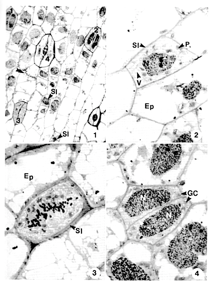
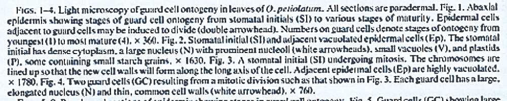
Guard cells in Ophioglossum petiolatum leaves are initiated by a single division of a stomatal initial with no subsidiary cells being formed. The stomatal initials have few vacuoles, plastids with little starch, and a large nucleus with much heterochromatin and prominent nucleoli.
Young guard cells are similar cytologically to stomatal initials; their common cell walls are thin and traversed by plasmodesmata. Plasmodesmata are also present within guard cell and adjacent epidermal cell walls.
With increasing age, guard cells develop a lenticular thickening in the median portion of the common cell walls, larger vacuoles, and plastids with several starch grains. Numerous microtubules are present near the thickening wall which also has an electron-translucent region between the cell wall and the plasmalemma.
Many dictyosomes and mitochondria are present in the cytoplasm. Older guard cells become more vacuolated, with some of the vacuoles containing fibrillar or dense deposits.
Plastids become very large as a result of storing several large starch grains. In the thickened portion of the cell walls, the middle lamella and some of the adjacent cell wall appear to be degraded during stomatal pore formation.
Mature guard cells are highly vacuolated and have very thick electron-dense cell walls without plasmodesmata. Fluorescence microscopy following aniline blue staining shows that aniline blue positive materials are present around and within the thickened portion of the cell walls, at the junction of guard cells with epidermal cells, and as distinct spots in guard cell and epidermal cell walls during the early stages of ontogeny.
Mature guard cells, however, lack the distinct fluorescent spots, which are interpreted as plasmodesmata, in their walls.

You must be logged in to post a comment.